
Plate 17.337 Pons- Mesencephalic Junction
Ronald A. Bergman, Ph.D., Adel K. Afifi, M.D., Paul M. Heidger,
Jr., Ph.D.
Peer Review Status: Externally Peer Reviewed
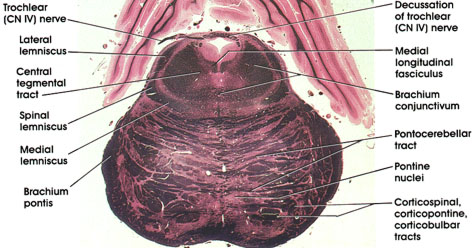
Human, 10% formalin, Pal-Weigert, 2.9 x.
Brachium conjunctivum: Massive outflow tract of the cerebellum. Fibers are seen just prior to and beginning decussation. Fibers project, after decussation, into the red nucleus and ventrolateral nucleus of the thalamus. Lesions in this tract result in a disorder of coordinated movement.
Pontocerebellar tract: The same structure seen at more caudal levels.
Corticospinal, corticopontine, corticobulbar tracts: The same structures seen at more caudal levels. Cut in cross section as they descend to lower caudal levels.
Pontine nuclei: Scattered between pontocerebellar fibers and the corticospinal, corticopontine, and corticobulbar tracts.
Brachium pontis: Axons of pontine nuclei on their way to the cerebellum.
Medial lemniscus: Continuation of the same structure seen at more caudal levels.
Spinal lemniscus: Continuation of the same structure seen at more caudal levels. Contains spinothalamic and spinotectal fibers.
Lateral lemniscus: Contains cochlear fibers. Located laterally and dorsally on its way to the inferior colliculus and medial geniculate body. Concerned with audition.
Trochlear (CN IV) nerve: Seen exiting from the dorsal aspect of the midbrain after decussating. The fourth cranial nerve supplies the superior oblique extraocular muscle. The only cranial nerve to decussate (cross) completely prior to leaving the neuraxis.
Central tegmental tract: Compact fiber bundle located medial to the brachium conjunctivum. Carries fibers from the midbrain tegmentum, red nucleus, and periaqueductal gray matter to the inferior olivary complex. Note change in position of this tract in more caudal levels.
Medial longitudinal fasciculus: Continuation of same structure seen at more rostral and more caudal levels.
Decussation of trochlear (CN IV) nerve: Axons of neurons in the trochlear nucleus seen decussating prior to exit from the dorsal surface of the neuraxis.
Next Page | Previous Page | Section Top | Title Page
Please send us comments by filling out our Comment Form.
All contents copyright © 1995-2024 the Author(s) and Michael P. D'Alessandro, M.D. All rights reserved.
"Anatomy Atlases", the Anatomy Atlases logo, and "A digital library of anatomy information" are all Trademarks of Michael P. D'Alessandro, M.D.
Anatomy Atlases is funded in whole by Michael P. D'Alessandro, M.D. Advertising is not accepted.
Your personal information remains confidential and is not sold, leased, or given to any third party be they reliable or not.
The information contained in Anatomy Atlases is not a substitute for the medical care and advice of your physician. There may be variations in treatment that your physician may recommend based on individual facts and circumstances.
URL: http://www.anatomyatlases.org/