
Plate 17.342 Diencephalon
Ronald A. Bergman, Ph.D., Adel K. Afifi, M.D., Paul M. Heidger,
Jr., Ph.D.
Peer Review Status: Externally Peer Reviewed
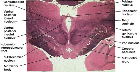
Human, 10% formalin, Pal-Weigert, 2.4 x.
Pulvinar nucleus: A thalamic nucleus. Belongs to the lateral group of thalamic nuclei. Has reciprocal connections with the medial and lateral geniculate bodies caudally and with the association parietal, temporal, and occipital cortices rostrally. Plays a role in several neural functions, including vision, audition, speech, and pain.
Lateral geniculate nucleus: A thalamic relay nucleus concerned with vision. Receives fibers from the optic tract and projects to the primary visual cortex (calcarine gyrus, area 17).
Red nucleus: So-called because of a pinkish color in the fresh state owing to its high vascularity. Links the cerebellum, cerebral cortex, and spinal cord.
Cerebral peduncle: A massive corticofugal fiber bundle. Continuous with internal capsule. Lesions here result in contralateral weakness or paralysis of the body, including the face.
Substantia nigra: Located dorsal to the cerebral peduncle. A mass of pigmented cells (melanin). This area is invariably the site of pathological changes associated with Parkinson's disease.
Mamillary body: A pair of spherical nuclear masses protruding from the ventral surface of the posterior hypothalamus. Receives fibers from the hippocampus via the fornix and is reciprocally connected with the anterior thalamic nuclei via the mamillothalamic tract. Concerned with memory function.
Subthalamic nucleus: Also known as corpus Luysii. Shaped like a biconcave lens. Receives fibers from and projects to the globus pallidus. Discrete lesions here result in an abnormal type of flinging movement known as ballism. Luys was a nineteenth-century French physician.
Habenulopeduncular tract: A fiber bundle coursing in the tegmenturn of the caudal diencephalon. Connects the habenular nuclei with the interpeduncular nucleus seen at more caudal levels. Note relation to red nucleus.
Ventral posterior medial nucleus and ventral posterior lateral nucleus: These two nuclei are collectively known as the ventral posterior thalamic nucleus or the ventrobasal complex. Belong to the lateral group of thalamic nuclei. A major relay station in the pathway of exteroceptive and proprioceptive impulses to the cortex. Receive fibers from the medial lemniscus, the spinothalamic tract, and the trigeminothalamic pathways. Reciprocally connected with the primary sensory cortex. The ventral posterior lateral nucleus receives the medial lemniscus and spinothalamic pathways, whereas the ventral posterior medial nucleus receives the trigeminothalamic pathway and taste sensations.
Centromedian nucleus: Belongs to the intralaminar group of thalamic nuclei. Almost completely surrounded by fibers of the internal medullary lamina.
Habenular nucleus: One of the nuclei of the epithalamus. Located in the caudal diencephalon dorsomedial to the thalamus. Receives the stria medullaris thalami and projects via the habenulopeduncular tract (fasciculus retroflexus of Meynert*) to the interpeduncular nucleus of the midbrain. The two habenular nuclei are connected by the habenular commissure. The habenular nuclei, part of a neural network that includes the limbic and olfactory systems, are concerned with mechanisms of emotion and behavior.
Third ventricle: Sandwiched between the two thalami.
*Meynert was a nineteenth-century Viennese neurologist.
Next Page | Previous Page | Section Top | Title Page
Please send us comments by filling out our Comment Form.
All contents copyright © 1995-2024 the Author(s) and Michael P. D'Alessandro, M.D. All rights reserved.
"Anatomy Atlases", the Anatomy Atlases logo, and "A digital library of anatomy information" are all Trademarks of Michael P. D'Alessandro, M.D.
Anatomy Atlases is funded in whole by Michael P. D'Alessandro, M.D. Advertising is not accepted.
Your personal information remains confidential and is not sold, leased, or given to any third party be they reliable or not.
The information contained in Anatomy Atlases is not a substitute for the medical care and advice of your physician. There may be variations in treatment that your physician may recommend based on individual facts and circumstances.
URL: http://www.anatomyatlases.org/