
Plate 17.345 Diencephalon
Ronald A. Bergman, Ph.D., Adel K. Afifi, M.D., Paul M. Heidger,
Jr., Ph.D.
Peer Review Status: Externally Peer Reviewed
Human, 10% formalin, Pal-Weigert, 2.0 x.
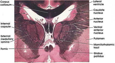
Corpus callosum: Massive fiber bundle connecting the two cerebral hemispheres. Important in interhemispheric transfer of information.
Caudate nucleus: Basal ganglia nucleus. Important in motor control.
Anterior (thalamic) nucleus: Located in rostral diencephalon. Receives the mamillothalarnic tract from the mamillary body and projects to the cingulate gyrus of the cerebral cortex. A component of the limbic system.
Ventral anterior (thalamic) nucleus: Medial to the internal capsule. Traversed by heavily myelinated fiber bundles. Receives fibers from the basal ganglia and is reciprocally connected with the motor cortex. Plays a role in motor control.
Putamen: One of the basal ganglia nuclei. Similar to the caudate nucleus in structure and connections. Along with the caudate it forms the neostriaturn. Lies ventral and lateral to the internal capsule.
Globus pallidus: Another of the basal ganglia nuclei. Receives fibers from caudate and putamen and projects to the thalamus (ventral anterior nucleus). Also reciprocally connected with the subthalamic nucleus.
Mamillothalamic tract: Seen entering the anterior nucleus of the thalamus. Axons of neurons in the mamillary body.
External medullary lamina: Between the internal capsule and ventral anterior nucleus. Defines the lateral boundary of the thalamus.
Internal capsule: The posterior limb, separates the basal ganglia (putamen and globus pallidus) from the thalamus.
Fornix: At this rostral level, the fornix bundle separates as the columns of the fornix arch ventrally and caudally en route to the mamillary body.
Next Page | Previous Page | Section Top | Title Page
Please send us comments by filling out our Comment Form.
All contents copyright © 1995-2024 the Author(s) and Michael P. D'Alessandro, M.D. All rights reserved.
"Anatomy Atlases", the Anatomy Atlases logo, and "A digital library of anatomy information" are all Trademarks of Michael P. D'Alessandro, M.D.
Anatomy Atlases is funded in whole by Michael P. D'Alessandro, M.D. Advertising is not accepted.
Your personal information remains confidential and is not sold, leased, or given to any third party be they reliable or not.
The information contained in Anatomy Atlases is not a substitute for the medical care and advice of your physician. There may be variations in treatment that your physician may recommend based on individual facts and circumstances.
URL: http://www.anatomyatlases.org/