
Plate 17.354 Thalamus and Basal Ganglia
Ronald A. Bergman, Ph.D., Adel K. Afifi, M.D., Paul M. Heidger,
Jr., Ph.D.
Peer Review Status: Externally Peer Reviewed
Human, 10% formalin, Weigert's hematoxylin (Loyez), 1 x.
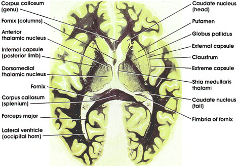
Corpus callosum: C-shaped bundle of heavily myelinated fibers. Connects the two hemispheres. Important in interhemispheric transfer of information. Section shows the genu (anterior) and splenium (posterior) parts of the corpus callosum.
Caudate nucleus: C-shaped mass of gray matter closely related to the lateral ventricle. A component of the basal ganglia and thus concerned with motor control. Section shows the head of the caudate (anterior) and the much smaller tail (posterior).
Putamen: A component of the basal ganglia. Plays a role in motor control. Medial to the external capsule and lateral to the caudate and globus pallidus.
Globus pallidus: One of the basal ganglia nuclei. Located medial to the putamen. The posterior limb of the internal capsule separates it from the thalamus. Traversed by heavily myelinated fiber bundles. Receives fibers from the caudate and putamen and projects to the thalamus (ventral anterior nucleus). Has reciprocal connections with the subthalamic nucleus. Plays a role in motor control.
External capsule: An efferent cortical fiber bundle. Lateral to the putamen.
Claustrum: A thin zone of gray substance located between the external and extreme capsules.
Extreme capsule: An efferent cortical fiber bundle. Situated lateral to the claustrum.
Stria medullaris thalami: A bundle of myelinated fibers connecting the septal nuclei with the habenular complex. Characteristically located dorsomedial to the thalamus.
Fimbria of fornix: Efferent fibers from the hippocampus. Continues as the body of the fornix.
Fornix: C-shaped, paired fiber system connecting the hippocampus with several brain regions, including the mamillary body, anterior thalamic nucleus, septal nuclei, and cingulate gyrus. The part of the fornix seen here represents the columns of the fornix.
Anterior thalamic nucleus: Belongs to the anterior group of thalamic nuclei. Has reciprocal connections with the mamillary body via the mamillothalamic tract and with the cingulate gyrus via the internal capsule. Considered part of the limbic system. Plays a role in emotional behavior and memory.
Internal capsule (posterior limb): Separates the putamen from the thalamus. Massive fiber bundle conveying impulses to and from the cerebral cortex. Lesions in internal capsule result in contralateral motor and sensory deficits.
Dorsomedial thalamic nucleus: Belongs to the medial group of thalamic nuclei. Most highly developed in man. Located medial to the internal medullary lamina. Has reciprocal connections with the prefrontal cortex and hypothalamus. Receives input from other thalamic nuclei. Concerned with affective behavior and memory.
Forceps major: Extension of the splenium of the corpus callosurn posteriorly into the occipital lobes.
Lateral ventricle: Extension of the cavity of the lateral ventricle into the occipital lobe (occipital horn).
Next Page | Previous Page | Section Top | Title Page
Please send us comments by filling out our Comment Form.
All contents copyright © 1995-2024 the Author(s) and Michael P. D'Alessandro, M.D. All rights reserved.
"Anatomy Atlases", the Anatomy Atlases logo, and "A digital library of anatomy information" are all Trademarks of Michael P. D'Alessandro, M.D.
Anatomy Atlases is funded in whole by Michael P. D'Alessandro, M.D. Advertising is not accepted.
Your personal information remains confidential and is not sold, leased, or given to any third party be they reliable or not.
The information contained in Anatomy Atlases is not a substitute for the medical care and advice of your physician. There may be variations in treatment that your physician may recommend based on individual facts and circumstances.
URL: http://www.anatomyatlases.org/