
Plate 17.362 Section through Cerebral Penduncle
Ronald A. Bergman, Ph.D., Adel K. Afifi, M.D., Paul M. Heidger,
Jr., Ph.D.
Peer Review Status: Externally Peer Reviewed
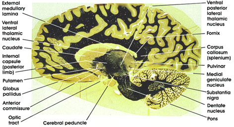
Human, 10% formalin, Weigert's hematoxylin (Loyez), 1 x.
Ventral lateral (thalamic) nucleus: One of the lateral group of thalmic nuclei. Characteristically traversed by heavily myelinated fiber bundles. Receives fibers from the cerebellum and is reciprocally connected with the primary motor cortex. Concerned with motor control.
Internal capsule (posterior limb): Contains fibers destined for and leaving the cerebral cortex. This part of the internal capsule separates the thalamus from the basal ganglia.
Caudate nucleus: One of the basal ganglia nuclei. Concerned with motor function. Note the similarity in appearance between caudate and putamen.
Globus pallidus: Another of the basal ganglia nuclei. Characterized by heavily myelinated bundles traversing it. The two components of globus pallidus, the outer and inner segments, are seen in this section. Note proximity to anterior commissure.
Putamen: One of the basal ganglia nuclei. Concerned with motor function.
Anterior commissure: Compact fiber bundle located in close proximity to globus pallidus. interconnects the olfactory bulbs and the temporal cortices.
Optic tract: Myelinated fiber bundle conveying visual impulses from the contralateral field of vision to the lateral geniculate nucleus and the pretectal area.
Pons: Note the characteristic ventral bulge known as the basis pontis.
Cerebral peduncle: Located ventral to the substantia nigra. Contains descending corticofugal fibers, including the corticospinal, corticobulbar, and corticopontine fiber systems. Lesions result in weakness (paresis) or paralysis of the contralateral half of the body, including the face.
Dentate nucleus: One of the deep cerebellar nuclei. Receives fibers from Purkinje neurons in the hemispheres of the cerebellum and projects to the thalamus (ventral lateral nucleus) and red nucleus via the dentatorubro thalamic system.
Substantia nigra: A large nuclear mass in the midbrain. Contains pigmented cells (melanin). Connected with the basal ganglia and thalamus. Important in motor control. This area is invariably the site of pathological changes associated with Parkinson's disease.
Medial geniculate nucleus: One of the thalamic nuclei. Concerned with audition. Receives auditory fibers from the brain stem and is reciprocally connected with the primary auditory cortex.
Pulvinar: One of the lateral group of thalamic nuclei. Reciprocally connected with the medial and lateral geniculate bodies caudally and the association parietal, temporal, and occipital cortices rostrally. Involved in several neural functions, including vision, audition, speech, and pain.
Corpus callosum (splenium): The caudal part of the corpus callosum. Important in transfer of visual information between the two hemispheres.
Fornix (crus): Continuation of the same system seen in other sections. The fimbria of the fornix arising from the hippocampus continues as the crus. Note relationship to splenium of corpus callosum.
Ventral posterior lateral (thalamic) nucleus: One of the lateral group of thalamic nuclei. Receives the medial lemniscus and the spinothalamic tract and projects to the primary somesthetic cortex in the postcentral gyrus.
External medullary lamina: A bundle of myelinated fibers separating the medial from the lateral group of thalamic nuclei.
Next Page | Previous Page | Section Top | Title Page
Please send us comments by filling out our Comment Form.
All contents copyright © 1995-2024 the Author(s) and Michael P. D'Alessandro, M.D. All rights reserved.
"Anatomy Atlases", the Anatomy Atlases logo, and "A digital library of anatomy information" are all Trademarks of Michael P. D'Alessandro, M.D.
Anatomy Atlases is funded in whole by Michael P. D'Alessandro, M.D. Advertising is not accepted.
Your personal information remains confidential and is not sold, leased, or given to any third party be they reliable or not.
The information contained in Anatomy Atlases is not a substitute for the medical care and advice of your physician. There may be variations in treatment that your physician may recommend based on individual facts and circumstances.
URL: http://www.anatomyatlases.org/