
Atlas of Human Anatomy
Translated by: Ronald A. Bergman, PhD and Adel K. Afifi, MD, MS
Peer
Review Status: Internally Peer Reviewed
Magnified View (via Quicktime VR)
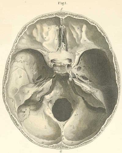
A) Frontal bone (orbital part).
B) Lesser wing of the sphenoid bone.
C) Greater wing of the sphenoid bone.
D) Squama of the temporal bone.
E) Petrous portion of the temporal bone.
F) Mastoid process of the temporal bone.
G) Occipital part of the occipital bone.
H) Basilar part of the occipital bone.
I) Lamina cribrosa of the ethmoid bone.
a) Cerebral ridge.
b) Digitate impressions.
c) Internal frontal spine, located in the longitudinal sulcus and it is attached
to the falx cerebri.
d) Crista galli is connected to the falx cerebri.
e) Foramina cribrosa allowing passage of olfactoria nerve.
f) Anterior clinoid process.
g) Optic foramen for the passage of the optic nerve and ophthalmic artery.
h) Middle clinoid process.
i) Sella turcica, the fossa for the hypophysis gland.
k) Posterior clinoid process.
l) Carotid sulcus and exit of the carotid canal.
m) Superior orbital fissure (passage for ophthalmic vein, and nn oculomotor,
trochlear, and abducens).
n) Foramen rotundum (for maxillary nerve).
o) Foramen ovale (for mandibular nerve).
p) Foramen spinosum (for middle meningeal artery, occasionally for the lesser
superficial petrosal nerve).
q) Canal or Hiatus for the greater petrosal nerve (passage for the nerve of
the pterygoid canal).
r) Internal auditory meatus (passage for acoustic artery, vein, and nerve and
the facial nerve).
s) Vestibular aqueduct.
t) Jugular foramen (passage for internal jugular vein, nn glossopharyngeal,
vagus, and spinal accessory).
u) Fossa for the medulla oblongata.
v) Clinoid process.
w) Anterior condyloid foramen (passage for hypoglossal nerve).
x) Posterior condyloid foramen (passage for Santorini emissary vein; often absent).
y) Mastoid foramen.
z) Foramen magnum (passage for medulla oblongata, accessory nerve, vertebral
artery and vein and spinal arteries).
a) Sigmoid fossa (a part of the transverse
sulcus).
b) Internal occipital spine (cruciate eminence).
g) Internal occipital ridge (for the falx
cerebelli).
d) Foramin cecum.
e) Ephippium with the clivus.
z) Foramen cecum.
h) Lingula.
q) Transverse sulcus.
i) Condyloid process of the occipital bone.
Please send us comments by filling out our Comment Form.
Anatomy Atlases is licensed under a Creative Commons Attribution-NonCommercial-ShareAlike 4.0 International License. 
"Anatomy Atlases", the Anatomy Atlases logo, and "A digital library of anatomy information" are all Trademarks of Michael P. D'Alessandro, M.D.
Anatomy Atlases is funded in whole by Michael P. D'Alessandro, M.D. Advertising is not accepted.
Your personal information remains confidential and is not sold, leased, or given to any third party be they reliable or not.
The information contained in Anatomy Atlases is not a substitute for the medical care and advice of your physician. There may be variations in treatment that your physician may recommend based on individual facts and circumstances.
URL: http://www.anatomyatlases.org/