
Atlas of Human Anatomy
Translated by: Ronald A. Bergman, PhD and Adel K. Afifi, MD, MS
Peer
Review Status: Internally Peer Reviewed
Magnified View (via Quicktime VR)
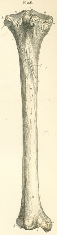
a) Medial condyle.
b) Lateral condyle.
c) Medial condylar cavity.
d) Lateral condylar cavity.
e) Intercondylar eminence (insertion for cruciate ligaments).
f) Superfical peroneal glenoid (socket) (for the fibular head).
g) Oblique line of the tibia (insertion site for mm popliteus and plantaris
and soleus.
h) Body, posterior surface.
i) Medial malleolus.
k) Fibular notch.
l) Groove for mm tibialis posterior and flexor digitorum longus.
Please send us comments by filling out our Comment Form.
Anatomy Atlases is licensed under a Creative Commons Attribution-NonCommercial-ShareAlike 4.0 International License. 
"Anatomy Atlases", the Anatomy Atlases logo, and "A digital library of anatomy information" are all Trademarks of Michael P. D'Alessandro, M.D.
Anatomy Atlases is funded in whole by Michael P. D'Alessandro, M.D. Advertising is not accepted.
Your personal information remains confidential and is not sold, leased, or given to any third party be they reliable or not.
The information contained in Anatomy Atlases is not a substitute for the medical care and advice of your physician. There may be variations in treatment that your physician may recommend based on individual facts and circumstances.
URL: http://www.anatomyatlases.org/