
Anatomy of First Aid: A Case Study Approach
Ronald Bergman, Ph.D.
Peer Review Status: Internally Peer Reviewed
A Machinist Mate is finishing work on a metal object when he suddenly feels scratchy, sharp pain in his eye and hurries to the medical corpsman for help.
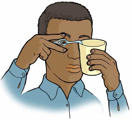
The corpsman first, tries to wash out the offending object with lukewarm water by splashing or flooding the eye with water, until blinking is not painful.
If this does not succeed in removing the object, blinking alone may flush the object from the eye. This may be very painful because the conjunctiva is richly supplied with pain nerve receptors.
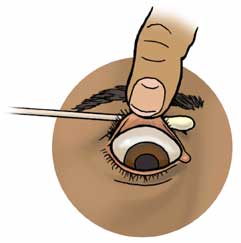
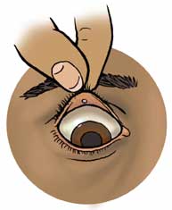
If this simple procedure also fails, the corpsman will examine the eye by lifting the eyelid from the eyeball. Several methods are shown in the illustrations.
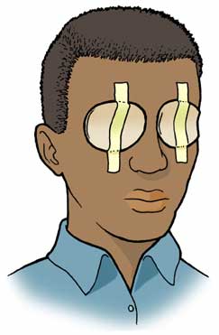
If the corpsman cannot find any object on the conjunctiva or cornea that would cause irritation, the object may be embedded in the eye. If this is the diagnosis, both eyes are covered with sterile or clean pads and taped in place. Medical advice or assistance will be sought.
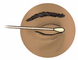
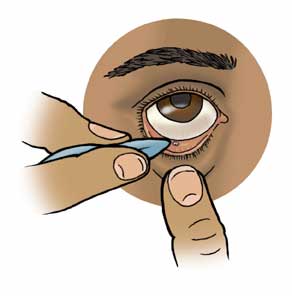
The eyelids and eye will be examined and if a foreign object is seen, it will be flushed directly to dislodge it or, with a clean moistened soft swab, the object is loosened and flushed, to remove the offending object. See illustrations.
If pain, tearing, or vision defects occur after removal of the foreign object or if the corpsman fails to find the cause of the problem he will seek medical advice and / or assistance.
Note: If any damage occurs to the eyeball, both eyes must be covered by sterile moist pads, and taped in place. Remember that the eyes are extensions of the brain and infections may ultimately involve the brain; this is to be avoided at all cost. A physician must treat a damaged eye, as soon as possible.
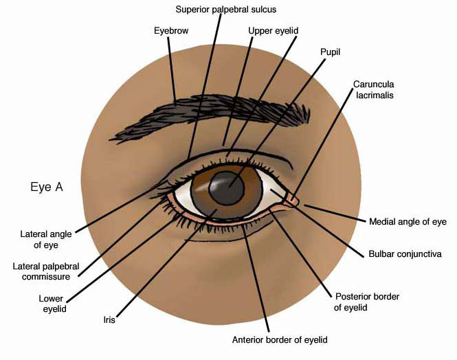
The anterior eye and eyelids.
EYE A. The eyeball is covered and protected anteriorly by two thin , movable eyelids (or palpebrae). The eyeball is also covered by a transparent mucous membrane (the conjunctiva), which is continuous along the inner surface of both eyelids (the palpebral conjunctiva).
At the medial angle of the eye a small piece of skin (the caruncula lacrimalis) is located that contains sebaceous and sweat glands.
The pupil is the circular opening in the iris. The size of the opening is controlled by the nervous system: at rest, the parasympathetic nervous system constricts the pupil and in danger, the sympathetic nervous system supplies the pupillary dilator muscle to enlarge the pupil.
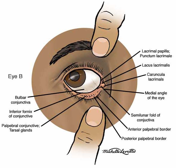
The lower eyelid and medial angle
EYE B. The lower eye lid has been pulled downward in order to expose its inner surface (i.e., the palpebral conjunctiva), as well as the medial angle (or medial canthus). The gaze is upward (superiorly) and outward (laterally).
The conjunctiva is very vascular and very sensitive. The inferior palpebral part and the bulbar part are continuous along a line of reflection (inferior conjunctival fornix). The line of reflection is also found between the eyeball and the upper eyelid (superior conjunctival fornix).
When the medial angle is enlarged, a pair of small openings (punctae lacrimali) are visible, located above and below the caruncula lacrimalis. These openings enter the lacrimal canals leading to the nasolacrimal duct and further, to the nose.
Please send us comments by filling out our Comment Form.
All contents copyright © 1995-2025 the Author(s) and Michael P. D'Alessandro, M.D. All rights reserved.
"Anatomy Atlases", the Anatomy Atlases logo, and "A digital library of anatomy information" are all Trademarks of Michael P. D'Alessandro, M.D.
Anatomy Atlases is funded in whole by Michael P. D'Alessandro, M.D. Advertising is not accepted.
Your personal information remains confidential and is not sold, leased, or given to any third party be they reliable or not.
The information contained in Anatomy Atlases is not a substitute for the medical care and advice of your physician. There may be variations in treatment that your physician may recommend based on individual facts and circumstances.
URL: http://www.anatomyatlases.org/