
Anatomy of First Aid: A Case Study Approach
Ronald Bergman, Ph.D.
Peer Review Status: Internally Peer Reviewed
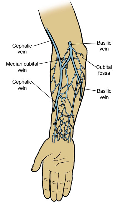
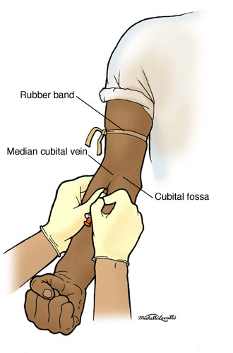
A hospital corpsman consulted her list of blood donor volunteers and asked one to donate a pint of O+ blood. It was possible that it might be needed for an emergency appendectomy being undertaken by the ship's surgeon while at sea. The volunteer Sailor was brought to sickbay and asked to lie down on the bed. The corpsman determined that the Sailor was healthy; his pulse, temperature and blood pressure were of normal values. She then tied a rubber band around the Sailor's arm, above the elbow, tight enough to stop superficial venous blood flow but not enough to prevent arterial blood flow. The cubital fossa (anterior surface of the elbow) was palpated and the median cubital vein was readily located (see illustrations), facilitated by the Sailor repeatedly making a fist. The corpsman knew that there were several large veins available in the region of the cubital fossa that she could use for venipuncture. She was aware that there is considerable normal variation in the pattern of veins in the arm and this is usually of no consequence. The corpsman then sponged clean the cubital fossa with alcohol and dried it with a sterile gauze pad. She inserted the IV catheter through the skin at an angle of about 45 degrees until she felt the needle enter the vein (by a slight decrease of resistance), then she decreased the angle of the syringe to about 10 to 20 degrees and advanced it slightly. Blood filled the lower part of the catheter reassuring the corpsman that she was indeed inside the vein. The plastic sleeve of the IV catheter was advanced over the catheter needle into the vein. The pressure band was then released. A blood collection bag was connected to the hypodermic needle and the hypodermic needle was carefully taped to the skin to prevent it from becoming dislodged. The corpsman had several types of catheter needles to select from but used the simplest one in this case.


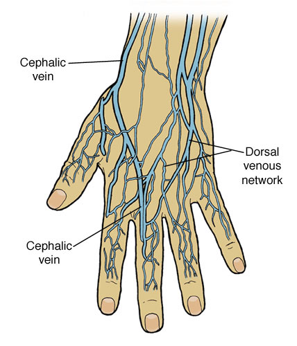
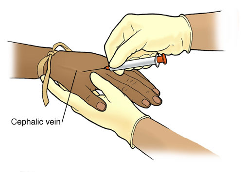
The back of the hand (the dorsum of the hand) is also available for venipuncture or IV insertion and here the veins are usually clearly seen. They are not tightly bound to surrounding tissues, hence they move and are deceptively easy to penetrate. If they are held in place by a finger, penetration is facilitated. Instead of the rubber band being applied around the arm when the back of the hand is selected for venipuncture, it is placed around the lower forearm above the wrist.


Please send us comments by filling out our Comment Form.
All contents copyright © 1995-2025 the Author(s) and Michael P. D'Alessandro, M.D. All rights reserved.
"Anatomy Atlases", the Anatomy Atlases logo, and "A digital library of anatomy information" are all Trademarks of Michael P. D'Alessandro, M.D.
Anatomy Atlases is funded in whole by Michael P. D'Alessandro, M.D. Advertising is not accepted.
Your personal information remains confidential and is not sold, leased, or given to any third party be they reliable or not.
The information contained in Anatomy Atlases is not a substitute for the medical care and advice of your physician. There may be variations in treatment that your physician may recommend based on individual facts and circumstances.
URL: http://www.anatomyatlases.org/