
Atlas of Human Anatomy in Cross Section: Section 1. Head and Neck
Ronald A. Bergman, Ph.D., Adel K. Afifi, M.D., Jean J. Jew, M.D., and Paul
C. Reimann, B.S.
Peer Review Status: Externally Peer Reviewed
|
Upper Left Quadrant |
Lower Left Quadrant |
Lower Right Quadrant |
Upper Right Quadrant |
|
1. Callosomarginal br. of anterior cerebral a. |
7. Middle cerebral a., br. |
11. Superior sagittal sinus |
16. Postcentral gyrus |
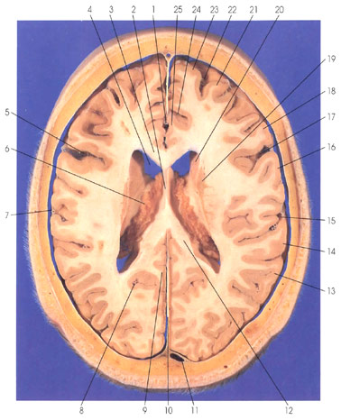
This is a section (looking down) through the body (6) and frontal (anterior) horn (4) of the lateral ventricle. The falx cerebri (10) is seen in the interhemispheric fissure. The superior sagittal sinus (11) is seen within a dural fold. The choroid plexus (6) is seen within the body of the lateral ventricle. The septum pellucidum (2) forms a partition between the two lateral ventricles. In the lateral wall of the frontal (anterior) horn (4) of the lateral ventricle is the head of the caudate nucleus (20). Lateral and inferior to the caudate nucleus is the putamen (19). The caudate and putamen constitute the striatum of the basal ganglia. The two hemispheres are connected by the corpus callosum (3, 12). The cingulate gyrus (25) is seen anterior and dorsal to the corpus callosum (3) on the medial surface of the hemisphere. The cingulate sulcus (24) delineates the boundary of the cingulate gyrus (25). Within the interhemispheric fissure rostrally is the callosomarginal branch ( 1 ) of the anterior cerebral artery. The central (rolandic) sulcus (5, 17) separates the frontal and parietal lobes. Rostral to the central (rolandic) sulcus (5, 17) is the precentral gyrus (18), which is the primary motor cortex. In the frontal lobe are also seen the superior (23) and middle (21) frontal gyri separated by the superior frontal sulcus (22). Caudal to the central (rolandic) sulcus (5, 17) is the postcentral gyrus (16), the primary somatosensory (somesthetic) cortex. The lateral (sylvian) fissure (15) is seen with branches of the middle cerebral artery (7). The supramarginal (14) and angular (13) gyri of the inferior parietal lobe are seen. The parietooccipital sulcus (8) separates the parietal and occipital lobes on their medial surface. The parietooccipital artery is seen within the sulcus (8). The precuneus gyrus (9) is seen rostral to the parietooccipital sulcus (8) in the parietal lobe.
Next Page | Previous Page | Section Top | Title Page
Please send us comments by filling out our Comment Form.
Anatomy Atlases is licensed under a Creative Commons Attribution-NonCommercial-ShareAlike 4.0 International License. 
"Anatomy Atlases", the Anatomy Atlases logo, and "A digital library of anatomy information" are all Trademarks of Michael P. D'Alessandro, M.D.
Anatomy Atlases is funded in whole by Michael P. D'Alessandro, M.D. Advertising is not accepted.
Your personal information remains confidential and is not sold, leased, or given to any third party be they reliable or not.
The information contained in Anatomy Atlases is not a substitute for the medical care and advice of your physician. There may be variations in treatment that your physician may recommend based on individual facts and circumstances.
URL: http://www.anatomyatlases.org/