
Atlas of Human Anatomy in Cross Section: Section 1. Head and Neck
Ronald A. Bergman, Ph.D., Adel K. Afifi, M.D., Jean J. Jew, M.D., and Paul
C. Reimann, B.S.
Peer Review Status: Externally Peer Reviewed
|
Upper Left Quadrant |
Lower Left Quadrant |
Lower Right Quadrant |
Upper Right Quadrant |
|
1. Brain, frontal lobe in anterior cranial fossa |
11. External auditory meatus |
24. Cerebellar tonsil |
32. Middle cranial fossa |
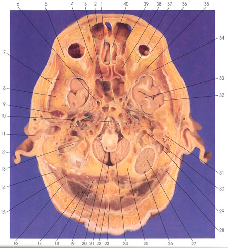
This is a section through the base of the cranial cavity. Rostrally in the midline is the crista galli (40). On one side of the crista galli (40) are the anterior (39) and posterior (38) ethmoid sinuses, and on the other is a frontal lobe (1) gyrus. Rostral to the anterior cranial fossa (1) is the frontal air sinus (2), and caudal to the posterior ethmoid sinus (38) is the spheroid sinus (3). Within the orbital cavity are the medial (4) and lateral (35) rectus muscles, orbital fat (5), the sclera (36), and optic nerve (37). Lateral and caudal to the orbital cavity is the greater wing of the spheroid bone (34). The temporal lobe (33) is seen in the middle cranial fossa (32). Within the posterior cranial fossa (26) are the hemispheres (27) and tonsils (24) of the cerebellum, the medulla oblongata (20) and the basilar artery (10). The tonsils of the cerebellum (24) and the medulla oblongata (20) are visualized through the foramen magnum (21) in the occipital bone (25). Major vascular structures seen in this section include the internal carotid artery (6), internal jugular vein (30), and the sigmoid sinus (13, 29). Lateral to the internal jugular vein (30) are the mastoid air cells (31). Bony landmarks in this section include the occipital (25) and temporal (8) bones, the greater wing of the sphenoid bone (34), and the condyloid process of the mandible (9). The pinna (12) and external auditory meatus (11) are seen. Outside the cranial cavity, the following muscles, tendons, and ligaments are seen: splenius capitis (28), trapezius (22), rectus capitis posterior minor (19) and major (18), semispinalis capitis (17), splenius capitis (16), rectus capitis lateralis (15), sternocleidomastoid tendon (14), ligamentum nuchae (23), and temporalis tendon (7).
Next Page | Previous Page | Section Top | Title Page
Please send us comments by filling out our Comment Form.
Anatomy Atlases is licensed under a Creative Commons Attribution-NonCommercial-ShareAlike 4.0 International License. 
"Anatomy Atlases", the Anatomy Atlases logo, and "A digital library of anatomy information" are all Trademarks of Michael P. D'Alessandro, M.D.
Anatomy Atlases is funded in whole by Michael P. D'Alessandro, M.D. Advertising is not accepted.
Your personal information remains confidential and is not sold, leased, or given to any third party be they reliable or not.
The information contained in Anatomy Atlases is not a substitute for the medical care and advice of your physician. There may be variations in treatment that your physician may recommend based on individual facts and circumstances.
URL: http://www.anatomyatlases.org/