
Atlas of Human Anatomy in Cross Section: Section 1. Head and Neck
Ronald A. Bergman, Ph.D., Adel K. Afifi, M.D., Jean J. Jew, M.D., and Paul
C. Reimann, B.S.
Peer Review Status: Externally Peer Reviewed
|
Upper Left Quadrant |
Lower Left Quadrant |
Lower Right Quadrant |
Upper Right Quadrant |
|
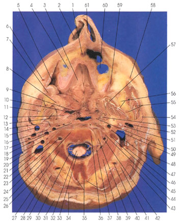
1. Incisive canal, posterior nasal septal a., and nasopalatine nerve |
12. Palatine tonsil |
36. Tendon m. semispinalis cervicis and vertebra, second cervical inferior
articular process |
55. Mandibular canal, inferior alveolar a. and nerve, and masseter m. |
This section passes through the nares (2), maxillary bone (4) and its incisive canal (1), pharynx (10), mandible (11, 56) and its mandibular canal (9, 55), and the second cervical vertebra (25, 27, 35, 36).
The superior pharyngeal constrictor muscle (10, 17) is thin and pale. Its fibers pass dorsally from an extensive anterior attachment to insert on the pharyngeal raphe, the pharyngeal tubercle of the occipital bone. The more inferior fibers arch distally deep to the middle constrictor muscle. The muscle has four sites of origin: from the dorsal margin of the medial pterygoid lamina and its hamular process, from the pterygomandibular raphe, from the posterior one fifth of the mylohyoid line of the mandible, and from the side of the root of the tongue deep to the hyoglossus muscle. It is innervated by the vagus nerve (CN 10) and it constricts the pharynx.
Note the bifid spinous process of the second cervical vertebra (35). The spinous processes of cervical vertebrae 2, 3, 4, and 5 usually possess bifid spines. The sixth and seventh show a tendency to bifurcate and usually present two small lateral tubercles. Occasionally all the cervical spines except the second are nonbifid. The only cervical vertebra that possesses a bifid spine in all races of man is the second.
Semispinalis cervicis arises from the transverse processes of the upper thoracic vertebrae and insert on the spinous processes of the second (36) through the fifth cervical vertebrae.
The suboccipital inferior oblique muscle (38) arises from the spine of the second cervical vertebra (axis) (35) and inserts onto the tip of the transverse process of the first cervical vertebra (atlas) (45). All four suboccipital muscles are innervated by the posterior primary ramus of the suboccipital (first cervical) nerve.
Next Page | Previous Page | Section Top | Title Page
Please send us comments by filling out our Comment Form.
Anatomy Atlases is licensed under a Creative Commons Attribution-NonCommercial-ShareAlike 4.0 International License. 
"Anatomy Atlases", the Anatomy Atlases logo, and "A digital library of anatomy information" are all Trademarks of Michael P. D'Alessandro, M.D.
Anatomy Atlases is funded in whole by Michael P. D'Alessandro, M.D. Advertising is not accepted.
Your personal information remains confidential and is not sold, leased, or given to any third party be they reliable or not.
The information contained in Anatomy Atlases is not a substitute for the medical care and advice of your physician. There may be variations in treatment that your physician may recommend based on individual facts and circumstances.
URL: http://www.anatomyatlases.org/