
Atlas of Human Anatomy in Cross Section: Section 3. Middle Thorax (Heart and Lungs)
Ronald A. Bergman, Ph.D., Adel K. Afifi, M.D., Jean J. Jew, M.D., and Paul
C. Reimann, B.S.
Peer Review Status: Externally Peer Reviewed
|
Upper Left Quadrant |
Lower Left Quadrant |
Lower Right Quadrant |
Upper Right Quadrant |
|
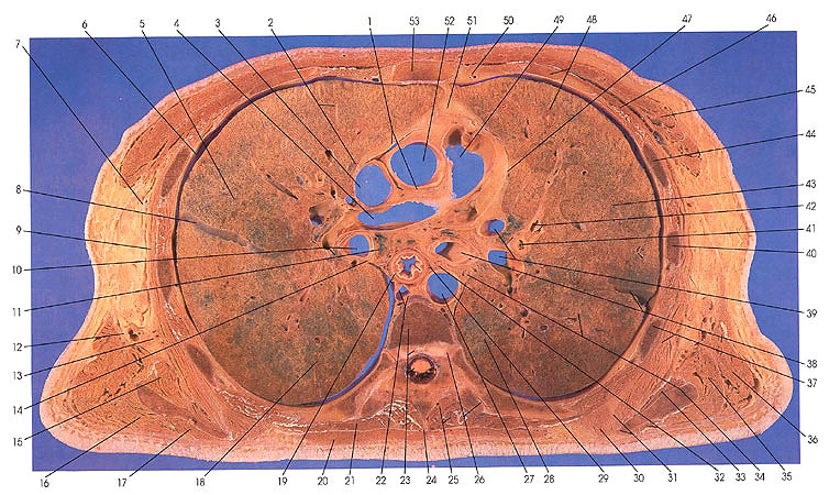
1. Pericardial cavity and transverse pericardial sinus |
9. Serratus anterior m. |
23. Spinal cord and disk, T6, and vertebral body, T6 |
40. Rib 5 |
This section passes through the sixth thoracic vertebra and a portion of the sixth intervertebral disk (23). It cuts the superior and transverse costal joints (26), ribs 7 (26) 6 (37), 5 (40), 4 (44), and 3 (48), and the sternum (53).
On the left side, the pulmonary artery (49) and its branches (39), the descending aorta (27), internal thoracic artery and veins (50), and the pericardiacophrenic artery (47) are seen. The phrenic nerve (47), left vagus nerve (34), left recurrent laryngeal nerve (32) esophageal plexus of autonomic nerves (29), and the spinal cord are important nervous tissue elements in this section. The left mainstem bronchus (38) and the secondary (41) and tertiary apical and posterior segmental (42) bronchi are identified. The lung left upper lobe, upper division, anterior (48) and apical posterior (43) and the left lower lobe, superior (28, 34) bronchopulmonary segments are shown. On the anterior thoracic wall, pectoralis major (45) and minor (46) muscles can be identified; on the lateral wall, serratus anterior (37) and latissimus dorsi (36) muscles are indicated; and on the posterior wall, teres major
(35), subscapularis (33), rhomboideus major (30), semispinalis thoracis (25), and multifidus (25) muscles are also labeled.
In the middle, the ascending aorta (52), right pulmonary artery (4), the descending aorta (27), the thoracic duct and azygos vein (22), and the esophagus (19) are found.
On the right side, the pulmonary artery (4), superior vena cave (3), lateral thoracic artery (7, 13), and the pericardiacophrenic artery (2) are seen. The right vagus (14), long thoracic (13), thoracodorsal (12), and the right phrenic (2) nerves are accounted for. The right lung upper lobe, superior bronchopulmonary segment (18); oblique interlobar fissure (8), and right upper lobe, posterior (5) and anterior (2) bronchopulmonary segments are identified. The trapezius (20), longissimus dorsi (21), infraspinatus (17), teres major (16), subscapularis (15), and serratus anterior (9) are also identified.
A mediastinal Iymph node is identified. The pericardial cavity and the transverse pericardial sinus (1) are seen.
Next Page | Previous Page | Section Top | Title Page
Please send us comments by filling out our Comment Form.
Anatomy Atlases is licensed under a Creative Commons Attribution-NonCommercial-ShareAlike 4.0 International License. 
"Anatomy Atlases", the Anatomy Atlases logo, and "A digital library of anatomy information" are all Trademarks of Michael P. D'Alessandro, M.D.
Anatomy Atlases is funded in whole by Michael P. D'Alessandro, M.D. Advertising is not accepted.
Your personal information remains confidential and is not sold, leased, or given to any third party be they reliable or not.
The information contained in Anatomy Atlases is not a substitute for the medical care and advice of your physician. There may be variations in treatment that your physician may recommend based on individual facts and circumstances.
URL: http://www.anatomyatlases.org/