
Atlas of Human Anatomy in Cross Section: Section 3. Middle Thorax (Heart and Lungs)
Ronald A. Bergman, Ph.D., Adel K. Afifi, M.D., Jean J. Jew, M.D., and Paul
C. Reimann, B.S.
Peer Review Status: Externally Peer Reviewed
|
Upper Left Quadrant |
Lower Left Quadrant |
Lower Right Quadrant |
Upper Right Quadrant |
|
1. Right coronary a. |
13. Lung, right upper lobe, lateral segment |
29. Thoracic spinal cord |
45. Lung, left lower lobe, superior segment |
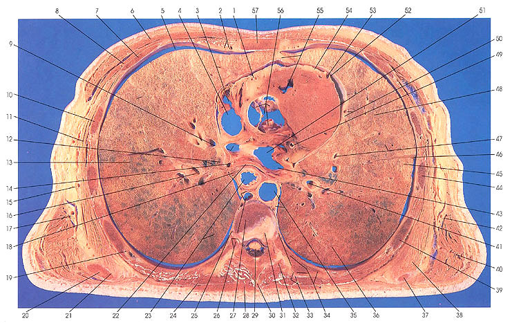
This section passes through the eighth thoracic vertebra (27) and grazes the eighth intervertebral disk (31). It cuts the eighth rib (34) and a costovertebral articulation or joint on the right side (26) and grazes the costotransverse joint (34) on the left side. It cuts the body of the sternum (57) in the midline.
The anterior mediastinum is represented by a small collection of fat (55).
The middle mediastinum contains the heart, and a number of its parts can be identified: semilunar valve components (tricuspid) and the aortic sinus (of Valsalva and of Petit) (56), the origin of the pulmonary artery (55), epicardium (54), anterior interventricular artery and vein (53), parietal pericardium (52), left atrium (51), left coronary artery (49), pulmonary vein (9, 14, 43), left atrial myocardium (22), superior vena cave (4), right atrium (3), and right coronary artery (1).
The posterior mediastinum contains the descending (thoracic) aorta (35), azygos vein and thoracic duct (26), the esophagus (23), mediastinal lymph nodes (18), and the aortic plexus and sympathetic trunk (36).
On the left side, the following bronchopulmonary segments have been indicated: left upper lobe, upper division, anterior (48); left upper lobe, upper division, apical posterior (47); left lower lobe, superior (45); and left lower lobe, superior (36).
On the right side, the following bronchopulmonary lymph segments have been indicated: right lower lobe, superior (23); right upper lobe, lateral (13); and right upper lobe, anterior (3).
Next Page | Previous Page | Section Top | Title Page
Please send us comments by filling out our Comment Form.
Anatomy Atlases is licensed under a Creative Commons Attribution-NonCommercial-ShareAlike 4.0 International License. 
"Anatomy Atlases", the Anatomy Atlases logo, and "A digital library of anatomy information" are all Trademarks of Michael P. D'Alessandro, M.D.
Anatomy Atlases is funded in whole by Michael P. D'Alessandro, M.D. Advertising is not accepted.
Your personal information remains confidential and is not sold, leased, or given to any third party be they reliable or not.
The information contained in Anatomy Atlases is not a substitute for the medical care and advice of your physician. There may be variations in treatment that your physician may recommend based on individual facts and circumstances.
URL: http://www.anatomyatlases.org/