
Atlas of Human Anatomy in Cross Section: Section 3. Middle Thorax (Heart and Lungs)
Ronald A. Bergman, Ph.D., Adel K. Afifi, M.D., Jean J. Jew, M.D., and Paul
C. Reimann, B.S.
Peer Review Status: Externally Peer Reviewed
|
Upper Left Quadrant |
Lower Left Quadrant |
Lower Right Quadrant |
Upper Right Quadrant |
|
1. Costomediastinal recess |
19. Intercostal neurovascular bundle |
36. Semispinalis dorsi and multifidus mm. |
50. Lung, left upper lobe, lower division (lingular) inferior segment,
and left pulmonary v. |
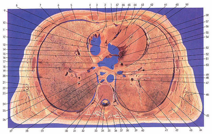
This section passes through the middle of the eighth thoracic vertebra (32), grazing the inferior vertebral notch (33) and transverse processes (38). The tip of the spinous process of T7 (35) is also seen. Ventrally, the section includes the body of the sternum (67) and its articulation (3) with the fourth rib (66) cartilage. The section cuts ribs 4 (6, 66), 5 (11), 6 (16), 7 (22), and 8 (41).
In the middle mediastinum, the right atrium (8) is seen at the entrance of the superior vena cave (14). The right inferior pulmonary vein (18) and the left inferior pulmonary vein (50) are joined to the left atrium (17). The foramen ovale (15) is seen but was found to be functionally closed (typical of about 25~ of hearts). The origin of the aorta (61) is seen
at this level, which is just inferior to the aortic (semilunar) valve.
A well defined transversus thoracis muscle (64) is seen. Teres major (43) and pectoralis major (7) and minor (9) make their last appearance at this level.
The upper and lower lobes of the left lung and the upper and lower lobes of the right lung and some bronchopulmonary segments (13, 20, 54, 65) are identified in this cut.
The nerve supply to latissimus dorsi (24, 46), the thoracodorsal nerve (23, 46); the nerve to serratus anterior (45), the long thoracic nerve (21); and the nerve supply to the intercostal muscles, the intercostal neurovascular bundle (19, 25), are seen.
The thoracic duct (29) is located slightly ventral and medial to the azygos vein (29).
Next Page | Previous Page | Section Top | Title Page
Please send us comments by filling out our Comment Form.
Anatomy Atlases is licensed under a Creative Commons Attribution-NonCommercial-ShareAlike 4.0 International License. 
"Anatomy Atlases", the Anatomy Atlases logo, and "A digital library of anatomy information" are all Trademarks of Michael P. D'Alessandro, M.D.
Anatomy Atlases is funded in whole by Michael P. D'Alessandro, M.D. Advertising is not accepted.
Your personal information remains confidential and is not sold, leased, or given to any third party be they reliable or not.
The information contained in Anatomy Atlases is not a substitute for the medical care and advice of your physician. There may be variations in treatment that your physician may recommend based on individual facts and circumstances.
URL: http://www.anatomyatlases.org/