
Atlas of Human Anatomy in Cross Section: Section 3. Middle Thorax (Heart and Lungs)
Ronald A. Bergman, Ph.D., Adel K. Afifi, M.D., Jean J. Jew, M.D., and Paul
C. Reimann, B.S.
Peer Review Status: Externally Peer Reviewed
|
Upper Left Quadrant |
Lower Left Quadrant |
Lower Right Quadrant |
Upper Right Quadrant |
|
1. Rib 5 and heart, right atrium |
9. Rib 7 |
25. Spinous process, T8 |
35. Lung, left lower lobe, anterior medial basal segment |
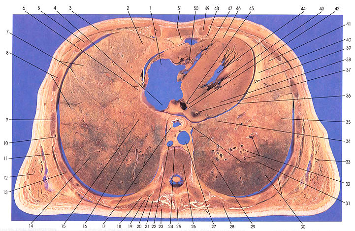
This section passes through the ninth thoracic vertebra (26), costotransverse articulation (21) and joint (20), the spinous process of the eighth thoracic vertebra (25), and, anteriorly, the body of the sternum (51). It cuts ribs 9 (18), 8 (13), 7 (9), 6 (8), and 5 (1, 5, 49).
In the middle mediastinum, the right atrioventricular valve (tricuspid) (50), right ventricle (47), anterior interventricular branch of the left coronary (left anterior descending) artery (46), coronary sinus (45), coronary sulcus (42), pericardial fat (40), left ventricular myocardium (38), pericardiacophrenic artery and phrenic nerve (39), great cardiac vein and circumflex branch of the left coronary artery (37), and the pericardial cavity (36) are seen.
On the left side, the left lung bronchopulmonary segments seen in this section include the left upper lobe, lower division (lingular), superior (41); left lower lobe, anterior medial basal (35); left upper lobe, lower division (lingular), inferior (34); left lower lobe, anterior medial basal (31); and the left lower lobe, lateral basal (30).
On the right side, the bronchopulmonary segments found include the right lower lobe, posterior basal (17); right middle lobe, medial (16); right middle lobe, medial basal (15); right lower lobe, lateral basal (14); right lower lobe, anterior basal (8); and the right middle lobe, lateral (7) and medial (2).
The esophagus (17), azygos vein (22), thoracic aorta (27), and the esophageal plexus of the vagus nerve (31) are seen. The trapezius muscle (23, 27) makes its last appearance on the left side in this section.
Next Page | Previous Page | Section Top | Title Page
Please send us comments by filling out our Comment Form.
Anatomy Atlases is licensed under a Creative Commons Attribution-NonCommercial-ShareAlike 4.0 International License. 
"Anatomy Atlases", the Anatomy Atlases logo, and "A digital library of anatomy information" are all Trademarks of Michael P. D'Alessandro, M.D.
Anatomy Atlases is funded in whole by Michael P. D'Alessandro, M.D. Advertising is not accepted.
Your personal information remains confidential and is not sold, leased, or given to any third party be they reliable or not.
The information contained in Anatomy Atlases is not a substitute for the medical care and advice of your physician. There may be variations in treatment that your physician may recommend based on individual facts and circumstances.
URL: http://www.anatomyatlases.org/