
Atlas of Human Anatomy in Cross Section: Section 5. Lower Thorax (Lungs) and Abdomen
Ronald A. Bergman, Ph.D., Adel K. Afifi, M.D., Jean J. Jew, M.D., and Paul
C. Reimann, B.S.
Peer Review Status: Externally Peer Reviewed
|
Upper Left Quadrant |
Lower Left Quadrant |
Lower Right Quadrant |
Upper Right Quadrant |
|
1. Mesenteric Iymph node |
10. Rib 10 |
26. Spinous process, L2 |
49. Descending colon |
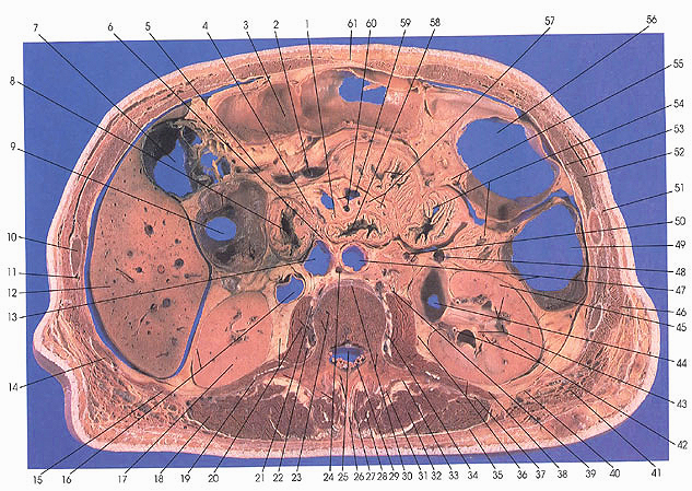
This section passes through the body of the second lumbar vertebra (23), and its pedicle (30), mamillary (24), spinous (26), and transverse (21) processes.
On the left side, the transverse (56) and descending colon (49) are seen along with various sections of the jejunum (54). In relation to the descending colon note the left colic artery (55), which arises from the inferior mesenteric artery and supplies the descending colon. The left colic vein is also seen. The left kidney is cut through the renal sinus and calix (43). Note that the renal cortex (45), medulla (44), and pyramid (44) can be readily differentiated from each other. The dilated proximal end of the ureter or pelvis (44) and renal arterial branches (42) are also identified. The anterior (47) and posterior (36) renal fascia, the perirenal capsular fat (20), the renal capsule (16), and the lumbar (37), psoas (36), and transversalis (40) fascia can all be seen.
In the middle of the section, the transverse colon (3), a coil of jejunum (57), jejunal arteries (57, 60), the superior mesenteric artery with a jejunal branch (60), and the superior mesenteric vein (59) can be identified.
Adjacent to the vertebral body, the abdominal aorta (47), inferior vena cave (13), sympathetic nerve trunk (39), and the left renal (retroaortic) vein (24) are seen.
On the right side, the right lobe of the liver (12) is diminishing in size, although the gallbladder (7) remains largely dilated. The ascending colon (9) is at the hepatic flexure; the duodenum (8) is taking a downward (posterior) course and is, in this section, immediately adjacent to the ascending colon (8, 9).
Next Page | Previous Page | Section Top | Title Page
Please send us comments by filling out our Comment Form.
Anatomy Atlases is licensed under a Creative Commons Attribution-NonCommercial-ShareAlike 4.0 International License. 
"Anatomy Atlases", the Anatomy Atlases logo, and "A digital library of anatomy information" are all Trademarks of Michael P. D'Alessandro, M.D.
Anatomy Atlases is funded in whole by Michael P. D'Alessandro, M.D. Advertising is not accepted.
Your personal information remains confidential and is not sold, leased, or given to any third party be they reliable or not.
The information contained in Anatomy Atlases is not a substitute for the medical care and advice of your physician. There may be variations in treatment that your physician may recommend based on individual facts and circumstances.
URL: http://www.anatomyatlases.org/