
Atlas of Human Anatomy in Cross Section: Section 5. Lower Thorax (Lungs) and Abdomen
Ronald A. Bergman, Ph.D., Adel K. Afifi, M.D., Jean J. Jew, M.D., and Paul
C. Reimann, B.S.
Peer Review Status: Externally Peer Reviewed
|
Upper Left Quadrant |
Lower Left Quadrant |
Lower Right Quadrant |
Upper Right Quadrant |
|
1. Transverse mesocolon |
13. Liver, right lobe |
28. Spinous process, L2 |
47. Left gonadal a. and v. |
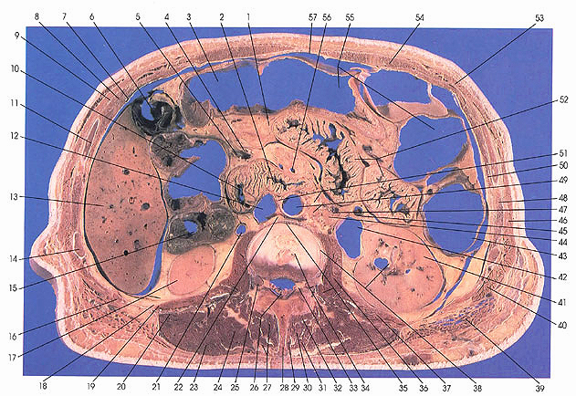
This section passes through the second lumbar intervertebral disk (between vertebrae L2 and L3). It grazes the inferior edge of the second lumbar vertebra and passes through the intervertebral foramina (25, 33), which contain dorsal root ganglia and some perineural fat. The spinous process of L2 (28) is also present. The section cuts ribs 12 (16), 11 (14), and 10 (11). Ribs make their last appearance in this section.
On the left side, the transverse (55) and descending colon (43) cover loops of jejunum (52). On the posterior abdominal wall, the left renal pelvis (42) and kidney (41) are seen. Among the muscles associated with the posterior abdominal wall the following may be seen: psoas major (36), quadratus lumborum (37), latissimus dorsi (39), serratus posterior inferior (40), and external oblique (46).
The middle of the section contains the transverse colon (55), the duodenal jejunal flexure (10), the horizontal part of the duodenum (2), the inferior vena cave (12), and the abdominal aorta (49). The retroaortic left renal vein (22) can be seen joining the inferior vena cave (12). Around the abdominal aorta, the aortic plexus of nerves (51) can be identified. The small intertransversarius muscle (35) can be identified below the psoas major muscle (36).
On the right side, the gallbladder (6) and right lobe of the liver can be seen. The gallbladder makes its last appearance in this section. Note the ascending colon (15), the lower pole of the right kidney (17), the capsular perirenal fat (17), and the pararenal fat (18) which is adjacent to, but outside, the renal fascia that forms the renal capsule.
The transversospinalis muscles are differentiated and well seen in this cut. They include the rotatores (32), semispinalis (31), multifidus (27, 30), and interspinalis (29) muscles.
Next Page | Previous Page | Section Top | Title Page
Please send us comments by filling out our Comment Form.
Anatomy Atlases is licensed under a Creative Commons Attribution-NonCommercial-ShareAlike 4.0 International License. 
"Anatomy Atlases", the Anatomy Atlases logo, and "A digital library of anatomy information" are all Trademarks of Michael P. D'Alessandro, M.D.
Anatomy Atlases is funded in whole by Michael P. D'Alessandro, M.D. Advertising is not accepted.
Your personal information remains confidential and is not sold, leased, or given to any third party be they reliable or not.
The information contained in Anatomy Atlases is not a substitute for the medical care and advice of your physician. There may be variations in treatment that your physician may recommend based on individual facts and circumstances.
URL: http://www.anatomyatlases.org/