
Atlas of Human Anatomy in Cross Section: Section 5. Lower Thorax (Lungs) and Abdomen
Ronald A. Bergman, Ph.D., Adel K. Afifi, M.D., Jean J. Jew, M.D., and Paul
C. Reimann, B.S.
Peer Review Status: Externally Peer Reviewed
|
Upper Left Quadrant |
Lower Left Quadrant |
Lower Right Quadrant |
Upper Right Quadrant |
|
1. Lymph node, lumbar |
15. Genitofemoral nerve |
31. Vertebral spinous process, L4 |
45. External abdominal oblique m. |
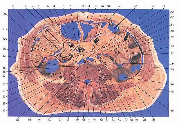
This section passes through the fourth lumbar vertebral body (26), the intervertebral foramina containing dorsal root ganglia and fourth lumbar nerves (28, 34), the arch (32) and spinous process of L4 (31), and, anteriorly, the umbilicus (62).
On the left side, the rectus abdominis muscle is seen within its sheath (57). Sections of the jejunum are still found on the left side adjacent to the descending colon (44). Superior mesenteric arterial and venous branches, including the middle colic and jejunal, are seen. The lumbar triangle is identified (42).
In the middle of the section, the transverse colon (56) and ileal arteries and veins (58) may be identified. The ileum (9) extends to the right side. At this level the bifurcation of the abdominal aorta into common iliac arteries (55) has almost been completed. The aortic plexus of nerves (53) is found. The union of the left common iliac vein with the inferior vena cave is taking place behind the aorta (23). Lumbar and mesocolic Iymph nodes (1, 8) are also found. The ureters (46, 13) are seen. Quadratus lumborum muscles (22, 40) are not seen below this section.
On the right side, coils of ileum (9) are cut. Ileocecal arteries and veins (7) are found. These branches of the superior mesenteric artery and vein (11) are found in relation to the ileum (9) and ascending (16) and transverse (3) colon. Ileocolic (5, 7), middle colic (2, 14) and right colic (21), gonadal (12), and superficial epigastric (6) blood vessels are also identified.
Next Page | Previous Page | Section Top | Title Page
Please send us comments by filling out our Comment Form.
Anatomy Atlases is licensed under a Creative Commons Attribution-NonCommercial-ShareAlike 4.0 International License. 
"Anatomy Atlases", the Anatomy Atlases logo, and "A digital library of anatomy information" are all Trademarks of Michael P. D'Alessandro, M.D.
Anatomy Atlases is funded in whole by Michael P. D'Alessandro, M.D. Advertising is not accepted.
Your personal information remains confidential and is not sold, leased, or given to any third party be they reliable or not.
The information contained in Anatomy Atlases is not a substitute for the medical care and advice of your physician. There may be variations in treatment that your physician may recommend based on individual facts and circumstances.
URL: http://www.anatomyatlases.org/