
Atlas of Human Anatomy in Cross Section: Section 5. Lower Thorax (Lungs) and Abdomen
Ronald A. Bergman, Ph.D., Adel K. Afifi, M.D., Jean J. Jew, M.D., and Paul
C. Reimann, B.S.
Peer Review Status: Externally Peer Reviewed
|
Upper Left Quadrant |
Lower Left Quadrant |
Lower Right Quadrant |
Upper Right Quadrant |
|
1. Lumbar lymph node |
10. Ascending colon and sympathetic trunk |
23. Internal vertebral venous plexus |
44. Jejunal a. and v. |
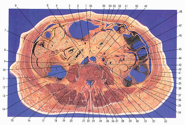
This section passes through the upper half of the fourth lumbar vertebra. The section crosses the transverse (29) and the superior articular process of L4 (27) and the inferior articular process of L3 (25).
On the left side, the transverse colon and its sacculations (46) can be seen. The colon can be identified by its size and the absence of mucosal folds and villi; the inner surface is smooth. The transverse colon joins the descending colon (34), and in the same region five cross sections of jejunum can be identified. Jejunal arteries and veins can be seen (44). The blood vessels found in this region arise from the superior mesenteric arteries and veins. The left ureter (43) and the left gonadal artery and vein (48) are identified.
In the middle of the section, a large sacculation of the transverse colon (3), middle colic arteries and veins (2), and lumbar Iymph nodes (1) are identified. The abdominal aorta (50) and inferior vena cave are seen before their bifurcation into iliac vessels, which are seen in the next section. An ascending lumbar vein (5) is seen.
On the right side, coils of ileum (6) are cut. Their mesentery contains ileal arteries and veins (7, 51), which are also branches of the superior mesenteric artery. The inferior edge of the right lobe of the liver (8) is seen. The liver makes its last appearance in this section. The ascending colon (10) is adjacent to the ileum (6), liver (8), and psoas major muscle (12). The right ureter (9) is seen.
Several important nerves are also seen: an intercostal nerve (38) between transversus abdominis (39) and internal oblique (40) muscles, the sympathetic trunk (10, 32), third and fourth lumbar nerves (30), the cauda equina nerve roots (24), iliohypogastric nerve (14), lateral (femoral) cutaneous nerve (of thigh) (13), and the genital branch of the genitofemoral nerve (12).
Next Page | Previous Page | Section Top | Title Page
Please send us comments by filling out our Comment Form.
Anatomy Atlases is licensed under a Creative Commons Attribution-NonCommercial-ShareAlike 4.0 International License. 
"Anatomy Atlases", the Anatomy Atlases logo, and "A digital library of anatomy information" are all Trademarks of Michael P. D'Alessandro, M.D.
Anatomy Atlases is funded in whole by Michael P. D'Alessandro, M.D. Advertising is not accepted.
Your personal information remains confidential and is not sold, leased, or given to any third party be they reliable or not.
The information contained in Anatomy Atlases is not a substitute for the medical care and advice of your physician. There may be variations in treatment that your physician may recommend based on individual facts and circumstances.
URL: http://www.anatomyatlases.org/