
Atlas of Human Anatomy in Cross Section: Section 5. Lower Thorax (Lungs) and Abdomen
Ronald A. Bergman, Ph.D., Adel K. Afifi, M.D., Jean J. Jew, M.D., and Paul
C. Reimann, B.S.
Peer Review Status: Externally Peer Reviewed
|
Upper Left Quadrant |
Lower Left Quadrant |
Lower Right Quadrant |
Upper Right Quadrant |
|
1. Lumbar Iymph nodes |
11. Right colic a. and w. |
25. Multifidus m. |
39. Gonadal a. and v. |
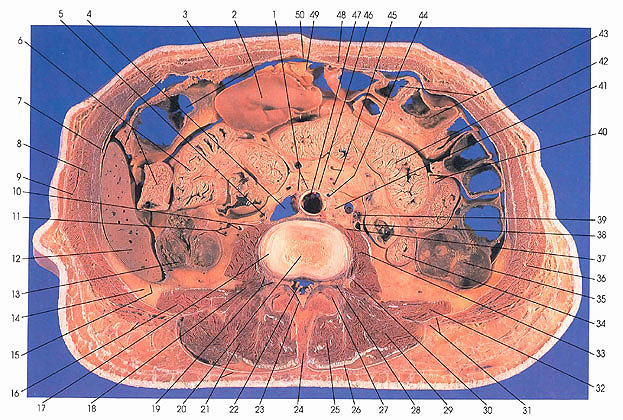
This section passes through the third lumbar intervertebral disk, where one may see the nucleus pulposus (20) and the anulus fibrosus (17). The spinous process of L3 (24) and the superior articular process of L4 (27) are also seen.
The three abdominal wall muscles-transversus abdominis (7), internal oblique (8), and external oblique (9)-are well developed in this cut. Between the internal and external oblique an intercostal, possibly T11, nerve can be found.
On the left side, the sacculations of the transverse colon (43), sections of ileum (42), appear, and the descending colon (35) is found in its usual left lower lateral position on the posterior abdominal wall. A few sections of jejunum (34) still remain at this level. The latissimus dorsi muscle is already aponeurotic and medial and does not appear on the left side. The thoracolumbar fascia (31) provides one point of origin of the abdominal oblique and transversus abdominis muscles. This area, devoid of an external covering of muscle, is known as the lumbar triangle (31).
In the middle of the section, an internal view of the colon (in general) reveals a smooth surface because the mucosal glands form pits and project downward rather than having luminal projections such as mucosal folds and villi, as is the case for the small intestine. The differentiation between the small bowel and the large bowel is usually readily made. Various primary and secondary branches of the abdominal aorta (47) are seen: gonadal (39), jejunal (40), inferior mesenteric (44), and right colic (11) arteries. The inferior vena cava (4) is adjacent to the aorta.
On the right side, the transverse colon (2) sacculations and the ascending colon (13) (which appears three sided) are seen. The three sided appearance is due to the restraining effect of the three tenia coli, bands of smooth muscle formed of the outer longitudinal layer of the tunica muscularis of the colon. The right lobe of the liver (12) is occupying a progressively smaller part of the right side of the abdomen. The peritoneum (15) is clearly marked on the right side separating the abdominal cavity from the subperitoneal fatty layer (14). Note the small intertransversarius muscle (18) beneath psoas major (17) and quadratus lumborum (16).
Next Page | Previous Page | Section Top | Title Page
Please send us comments by filling out our Comment Form.
Anatomy Atlases is licensed under a Creative Commons Attribution-NonCommercial-ShareAlike 4.0 International License. 
"Anatomy Atlases", the Anatomy Atlases logo, and "A digital library of anatomy information" are all Trademarks of Michael P. D'Alessandro, M.D.
Anatomy Atlases is funded in whole by Michael P. D'Alessandro, M.D. Advertising is not accepted.
Your personal information remains confidential and is not sold, leased, or given to any third party be they reliable or not.
The information contained in Anatomy Atlases is not a substitute for the medical care and advice of your physician. There may be variations in treatment that your physician may recommend based on individual facts and circumstances.
URL: http://www.anatomyatlases.org/