
Atlas of Human Anatomy in Cross Section: Section 6. Pelvis, Perineum, Hip, and Upper Thigh
Ronald A. Bergman, Ph.D., Adel K. Afifi, M.D., Jean J. Jew, M.D., and Paul
C. Reimann, B.S.
Peer Review Status: Externally Peer Reviewed
|
Upper Left Quadrant |
Lower Left Quadrant |
Lower Right Quadrant |
Upper Right Quadrant |
|
1. Median umbilical ligament |
14. Superior gluteal neurovascular bundle |
25. Anterior sacral plexus of w. |
38. Os ilium |
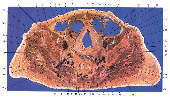
This section passes through the fourth sacral vertebra and sacral hiatus and the os ilium (38) at the sciatic notch (34).
The superior gluteal artery and vein (17, 32) can be seen traversing the greater sciatic foramen after supplying iliacus (42), piriformis (19, 30), and obturator internus (37) muscles and a nutrient artery to the ilium. Note the anterior sacral plexus of veins (25), which arise from the lateral sacral veins (which ultimately join the superior gluteal or internal iliac veins) and the median sacral vein. The median sacral vein communicates with the lateral sacral veins and with the anterior sacral plexus. These veins anastomose freely with neighboring lumbar, pelvic, and rectal veins. None of the veins have valves, hence they form a free communication between the systematic (caval) and portal systems of veins.
The obturator internus muscle (37) makes its first appearance in this cut.
The relationship between the peroneal (17) and tibial (19) nerves and piriformis muscle (19) is shown. The nerve to piriformis arises from the second or first and second sacral nerves, which contribute to the tibial nerve. The sciatic nerve is formed by the peroneal and tibial nerves within a common sheath that leaves the pelvis via the greater sciatic foremen, usually below the piriformis muscle.
Note the vermiform appendix (5) and its "mesentery." The appendix does not have a true mesentery; however, in about 90% of cases it is provided with a falciform fold of peritoneum, the mesoappendix, which is continuous with the dorsal (original left) surface of the mesentery of the ileum. In or near the free margin of the fold, the artery of the appendix, which is usually a branch of the ileocolic artery, may be found. The artery may also arise from the ileal, posterior cecal, or anterior cecal branch of the ileocecal artery. It usually passes dorsal to the ileum to enter the free border of the mesenteriolum of the appendix. The vessel may be doubled and it may also pass ventral to the ileum to supply the appendix.
Next Page | Previous Page | Section Top | Title Page
Please send us comments by filling out our Comment Form.
Anatomy Atlases is licensed under a Creative Commons Attribution-NonCommercial-ShareAlike 4.0 International License. 
"Anatomy Atlases", the Anatomy Atlases logo, and "A digital library of anatomy information" are all Trademarks of Michael P. D'Alessandro, M.D.
Anatomy Atlases is funded in whole by Michael P. D'Alessandro, M.D. Advertising is not accepted.
Your personal information remains confidential and is not sold, leased, or given to any third party be they reliable or not.
The information contained in Anatomy Atlases is not a substitute for the medical care and advice of your physician. There may be variations in treatment that your physician may recommend based on individual facts and circumstances.
URL: http://www.anatomyatlases.org/