
Atlas of Human Anatomy in Cross Section: Section 6. Pelvis, Perineum, Hip, and Upper Thigh
Ronald A. Bergman, Ph.D., Adel K. Afifi, M.D., Jean J. Jew, M.D., and Paul
C. Reimann, B.S.
Peer Review Status: Externally Peer Reviewed
|
Upper Left Quadrant |
Lower Left Quadrant |
Lower Right Quadrant |
Upper Right Quadrant |
|
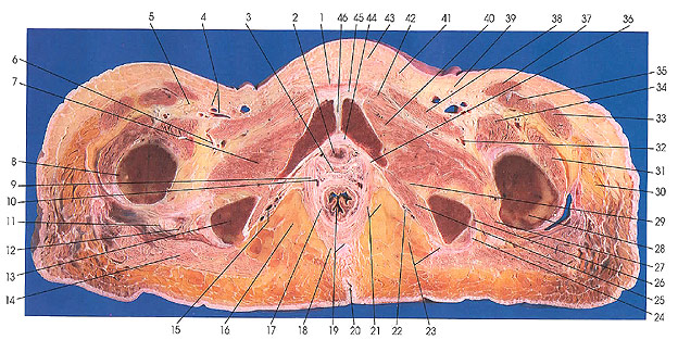
1. Pyramidalis m. |
9. Levator ani m. |
20. Anal crease |
31. Vastus lateralis m. |
This section passes through the pubic symphysis (46), urethra (2), vagina (3), anus (19), anal crease (20), and the femora (8, 28).
The levator ani muscles (9, 21) arise from the obturator fascia at the arcus tendineus (36).
Obturator internus muscle lies on the obturator membrane. It is covered by obturator fascia that is attached to the body of the pubis, the iliac portion of the terminal line, the ventral margin of the greater sciatic notch, the ischial spine, the sacrotuberous ligament and the falciform process of that ligament, and the ischial and pubic rami. Near the upper end of the obturator foremen the fascia is reflected over the obturator muscle and is attached to the obturator membrane. The upper part of the fascia is thickened near its middle part and forms a tendinous arch (36) for the attachment of the levator ani muscle (9, 21). Below the arch, the fascia forms the lateral boundary of the ischiorectal fossa (16, 21) and forms the pudendal canal (15, 22).
Next Page | Previous Page | Section Top | Title Page
Please send us comments by filling out our Comment Form.
Anatomy Atlases is licensed under a Creative Commons Attribution-NonCommercial-ShareAlike 4.0 International License. 
"Anatomy Atlases", the Anatomy Atlases logo, and "A digital library of anatomy information" are all Trademarks of Michael P. D'Alessandro, M.D.
Anatomy Atlases is funded in whole by Michael P. D'Alessandro, M.D. Advertising is not accepted.
Your personal information remains confidential and is not sold, leased, or given to any third party be they reliable or not.
The information contained in Anatomy Atlases is not a substitute for the medical care and advice of your physician. There may be variations in treatment that your physician may recommend based on individual facts and circumstances.
URL: http://www.anatomyatlases.org/