
Atlas of Human Anatomy in Cross Section: Section 6. Pelvis, Perineum, Hip, and Upper Thigh
Ronald A. Bergman, Ph.D., Adel K. Afifi, M.D., Jean J. Jew, M.D., and Paul
C. Reimann, B.S.
Peer Review Status: Externally Peer Reviewed
|
Upper Left Quadrant |
Lower Left Quadrant |
Lower Right Quadrant |
Upper Right Quadrant |
|
1. Mons pubis and labium majus |
13. Tendon m. fascia lata |
22. Anus |
32. iliopsoas m. |
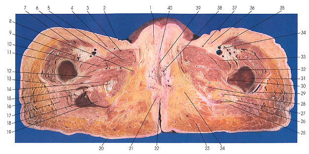
This section passes through the clitoris (40), vagina (39), anus (22), the femora (12, 31), and the lesser trochanter of the femur (30).
The muscles of the upper thigh dominate the section. Of the anterior group of muscles, those seen at this level include the sartorius (6) and quadriceps femoris (7, 11). They are innervated by the femoral nerve (34).
The medial group of muscles is represented by gracilis (8, 38), pectineus (33), adductor brevis (5, 10), adductor longus (3), adductor minimus (a part of adductor magnus) (12), and adductor magnus (14, 15, 19, 28). These muscles are innervated by the obturator nerve (8) except pectineus, which gets its nerve supply from both the femoral and obturator nerves (34), and adductor magnus, which gets its nerve supply from the sciatic. Adductor minimus is supplied by the nerve to quadratus femoris (lumbosacral trunk and first sacral nerve).
The posterior group of muscles is represented by semimembranosus (28), semitendinosus (26), and biceps femoris (26). These muscles are innervated by the tibial division of the sciatic nerve (16, 27) except the short head of the biceps. The short head is not seen at this level, but it is innervated by the peroneal nerve.
Next Page | Previous Page | Section Top | Title Page
Please send us comments by filling out our Comment Form.
Anatomy Atlases is licensed under a Creative Commons Attribution-NonCommercial-ShareAlike 4.0 International License. 
"Anatomy Atlases", the Anatomy Atlases logo, and "A digital library of anatomy information" are all Trademarks of Michael P. D'Alessandro, M.D.
Anatomy Atlases is funded in whole by Michael P. D'Alessandro, M.D. Advertising is not accepted.
Your personal information remains confidential and is not sold, leased, or given to any third party be they reliable or not.
The information contained in Anatomy Atlases is not a substitute for the medical care and advice of your physician. There may be variations in treatment that your physician may recommend based on individual facts and circumstances.
URL: http://www.anatomyatlases.org/