
Atlas of Human Anatomy in Cross Section: Section 7. Lower Limb
Ronald A. Bergman, Ph.D., Adel K. Afifi, M.D., Jean J. Jew, M.D., and Paul
C. Reimann, B.S.
Peer Review Status: Externally Peer Reviewed
|
Upper Left Quadrant |
Lower Left Quadrant |
Lower Right Quadrant |
Upper Right Quadrant |
|
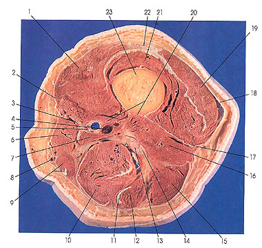
1. Vastus medialis m. |
4. Femoral a. and v. |
12. Semitendinosus m. |
18. Iliotibial tract (Maissiat's band) |
This section is three sections (3 cm) below the preceding section.
The rectus femoris has become tendinous (22), the quadriceps tendon. This tendon may be divided into three distinct layers. The superficial layer is usually formed by the rectus femoris, the intermediate layer by vastus lateralis (19) and vastus medialis (1), and the deep layer by vastus intermedius (21) muscle. Occasionally, vastus lateralis and medialis tendons may send tendinous fiber bundles crossing in front of the rectus tendon. The combined tendon of quadriceps attaches to the superior and medial borders of the patella, and in part in front and beyond the patella to join the patellar ligament. Some of the tendinous fibers of vastus lateralis and vastus medialis run on each side of the patella to the tibial condyles. These fibers are named the medial and lateral patellar ligaments. From the apex of the patella to the tibial tuberosity the quadriceps tendon continues as the patellar ligament.
The adductor canal (Hunter's' canal) (5) is identified in this section for the first time. The adductor canal extends through the middle third of the thigh. Beginning at the apex of the femoral triangle, it is bounded by the sartorius (6) ventrally, vastus medialis (1) medially, and adductor longus and magnus (7) dorsolaterally. It is roughly triangular in shape. The canal ends at the adductor hiatus (seen in the next cut). The canal carries the femoral artery (4) through the middle third of the thigh and contains, in addition, the femoral vein (4), nerve to vastus medialis muscle (20), and the saphenous nerve (3).
The iliotibial tract (Maissiat's' band) is identified.
(a)John Hunter (1728-1793) was a Scottish surgeon, anatomist, physiologist,
and pathologist. "
(2)Jacques Maissiat (1805-1878) was a French anatomist working in Parts. Next
Page | Previous Page | Section
Top | Title Page
Please send us comments by filling out our Comment Form.
Anatomy Atlases is licensed under a Creative Commons Attribution-NonCommercial-ShareAlike 4.0 International License. 
"Anatomy Atlases", the Anatomy Atlases logo, and "A digital library of anatomy information" are all Trademarks of Michael P. D'Alessandro, M.D.
Anatomy Atlases is funded in whole by Michael P. D'Alessandro, M.D. Advertising is not accepted.
Your personal information remains confidential and is not sold, leased, or given to any third party be they reliable or not.
The information contained in Anatomy Atlases is not a substitute for the medical care and advice of your physician. There may be variations in treatment that your physician may recommend based on individual facts and circumstances.
URL: http://www.anatomyatlases.org/