
Atlas of Human Anatomy in Cross Section: Section 7. Lower Limb
Ronald A. Bergman, Ph.D., Adel K. Afifi, M.D., Jean J. Jew, M.D., and Paul
C. Reimann, B.S.
Peer Review Status: Externally Peer Reviewed
|
Upper Left Quadrant |
Lower Left Quadrant |
Lower Right Quadrant |
Upper Right Quadrant |
|
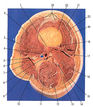
1. Medial lip of linea aspera of femur |
4. Medial intermuscular septum and popliteal a. and v. |
12. Semitendinosus m. and tendon |
17. Lateral intermuscular septum |
This section is three sections (3 cm) below the preceding one. The section cuts the adductor canal hiatus (8). It is at this point that the femoral artery and vein are renamed popliteal artery and vein (4). They will retain this name until the vessels reach the lower border of the popliteus muscle, after which they are renamed. The popliteal vessels terminate when they divide into the anterior and posterior branches: the anterior and posterior tibial artery and vein.
Note the reduction in size of the biceps femoris (long head) (15), semitendinosus (12), and gracilis (10) muscles. They lie beneath biceps femoris (short head) (16), semimembranosus (11), and sartorius (7) muscles, respectively.
Note the divergence of the lips of linea aspera (1, 18), with biceps femoris occupying the interval between the two lips. The divergence will continue and define a triangular surface of bone, the popliteal surface (planum popliteum).
The tibial nerve usually arises from lumbar nerves 4 and 5 and sacral nerves I and 2, and occasionally sacral 3 in addition. The common peroneal nerve arises from lumbar 4 and 5 and sacral nerves 1 and 2.
The posterior femoral cutaneous nerve (also known as the small sciatic nerve) arises from sacral nerves 1, 2, and 3.
Next Page | Previous Page | Section Top | Title Page
Please send us comments by filling out our Comment Form.
Anatomy Atlases is licensed under a Creative Commons Attribution-NonCommercial-ShareAlike 4.0 International License. 
"Anatomy Atlases", the Anatomy Atlases logo, and "A digital library of anatomy information" are all Trademarks of Michael P. D'Alessandro, M.D.
Anatomy Atlases is funded in whole by Michael P. D'Alessandro, M.D. Advertising is not accepted.
Your personal information remains confidential and is not sold, leased, or given to any third party be they reliable or not.
The information contained in Anatomy Atlases is not a substitute for the medical care and advice of your physician. There may be variations in treatment that your physician may recommend based on individual facts and circumstances.
URL: http://www.anatomyatlases.org/