
Atlas of Human Anatomy in Cross Section: Section 7. Lower Limb
Ronald A. Bergman, Ph.D., Adel K. Afifi, M.D., Jean J. Jew, M.D., and Paul
C. Reimann, B.S.
Peer Review Status: Externally Peer Reviewed
|
Upper Left Quadrant |
Lower Left Quadrant |
Lower Right Quadrant |
Upper Right Quadrant |
|
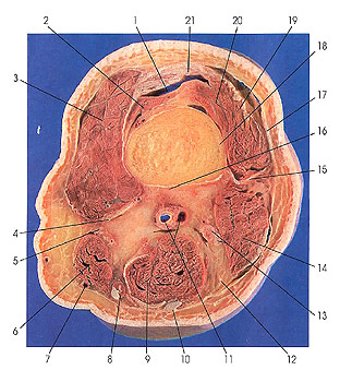
1. Suprapatellar bursa |
4. Tendon m. adductor magnus |
10. Tendon m. semitendinosus |
15. Lateral intermuscular septum |
This section is three sections (3 cm) below the preceding one.
This section passes through the popliteal surface (planum popliteum) formed by the divergence of the lateral and medial lips of linea aspera. In this section, the suprapatellar bursa (1) is seen for the first time. This bursa is located between the anterior surface of the lower end of the femur and the quadriceps tendon. It communicates with the joint cavity.
Vastus intermedius muscle (20) is composed of lamellae superimposed concentrically around the shaft of the femur. The deepest and most distal of these is called the articularis genu muscle (2). The fiber bundles of this layer are inserted into the capsule of the joint, or onto the superior margin of the patella, and ultimately, through the patellar ligament, onto the tuberosity of the tibia.
The saphenous nerve (5) is entirely sensory and supplies the skin on the medial side of the leg and foot via medial crural and infrapatellar branches.
The fasciae of the thigh and lower limb, in general, are well developed. The fascia lata encloses the muscles of the back of the hip and thigh and is especially strong on the lateral side, where it includes the longitudinal band of connective tissue fibers known as the iliotibial tract (17). From the fascia lata, strong intermuscular septa( 15) extend beneath the quadriceps group of muscles (3, 19, 20, 21) from the lateral and medial sides to join the periosteum of the femur.
Note that gracilis (8), semitendinosus (10), and adductor magnus (4) muscles are largely tendinous in this cut.
This cut ends the middle and lower thigh series. The next series of sections pass through the knee joint.
Next Page | Previous Page | Section Top | Title Page
Please send us comments by filling out our Comment Form.
Anatomy Atlases is licensed under a Creative Commons Attribution-NonCommercial-ShareAlike 4.0 International License. 
"Anatomy Atlases", the Anatomy Atlases logo, and "A digital library of anatomy information" are all Trademarks of Michael P. D'Alessandro, M.D.
Anatomy Atlases is funded in whole by Michael P. D'Alessandro, M.D. Advertising is not accepted.
Your personal information remains confidential and is not sold, leased, or given to any third party be they reliable or not.
The information contained in Anatomy Atlases is not a substitute for the medical care and advice of your physician. There may be variations in treatment that your physician may recommend based on individual facts and circumstances.
URL: http://www.anatomyatlases.org/