
Atlas of Human Anatomy in Cross Section: Section 7. Lower Limb
Ronald A. Bergman, Ph.D., Adel K. Afifi, M.D., Jean J. Jew, M.D., and Paul
C. Reimann, B.S.
Peer Review Status: Externally Peer Reviewed
|
Upper Left Quadrant |
Lower Left Quadrant |
Lower Right Quadrant |
Upper Right Quadrant |
|
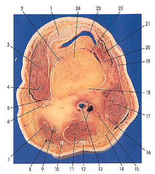
1. Tendon m. quadriceps femoris (quadriceps tendon) |
5. Superior medial genicular a. |
12. Tendon m. semitendinosus |
19. Iliotibial tract (Maissiat's band) |
This is the first section in the knee region. The suprapatellar bursa (23), which is continuous with the articular cavity (knee Joint), is seen. The femur (24) is cut in the supracondylar region. The vastus intermedius and articularis genu muscles are no longer present, but parts of vastus lateralis can be seen (21). Vastus medialis (3) is the largest muscle mass at this level, followed by biceps femoris (17). Note that semitendinosus (12) and gracilis are entirely tendinous.
The common peroneal (16) and tibial (15) nerves are seen closely associated with, and located between, biceps femoris (17) and the popliteal artery and vein (13).
Important elements of the blood supply of the knee Joint can be seen in this section. These arterial vessels are the superior lateral genicular (122); lateral femoral circumflex, descending branch (20); descending genicular, saphenous branch (9); superior medial genicular (2, 5); and the descending genicular, musculoarticular branch (4).
The deep articular plexus, around the knee joint, is formed by two medial and two lateral genicular branches of the popliteal, the descending genicular, the descending branch of the lateral femoral circumflex, the anterior and posterior tibial recurrent, the circumflex fibular, and the medial inferior genicular arteries.
The vastus lateralis (2 1), at this level, is supplied by the lateral femoral circumflex artery, descending branch (20); the vastus medialis (3) by the descending genicular artery, musculoarticular branch (4); and the sartorius (7) by the descending genicular artery, saphenous branch (9).
Next Page | Previous Page | Section Top | Title Page
Please send us comments by filling out our Comment Form.
Anatomy Atlases is licensed under a Creative Commons Attribution-NonCommercial-ShareAlike 4.0 International License. 
"Anatomy Atlases", the Anatomy Atlases logo, and "A digital library of anatomy information" are all Trademarks of Michael P. D'Alessandro, M.D.
Anatomy Atlases is funded in whole by Michael P. D'Alessandro, M.D. Advertising is not accepted.
Your personal information remains confidential and is not sold, leased, or given to any third party be they reliable or not.
The information contained in Anatomy Atlases is not a substitute for the medical care and advice of your physician. There may be variations in treatment that your physician may recommend based on individual facts and circumstances.
URL: http://www.anatomyatlases.org/