
Atlas of Human Anatomy in Cross Section: Section 7. Lower Limb
Ronald A. Bergman, Ph.D., Adel K. Afifi, M.D., Jean J. Jew, M.D., and Paul
C. Reimann, B.S.
Peer Review Status: Externally Peer Reviewed
|
Upper Left Quadrant |
Lower Left Quadrant |
Lower Right Quadrant |
Upper Right Quadrant |
|
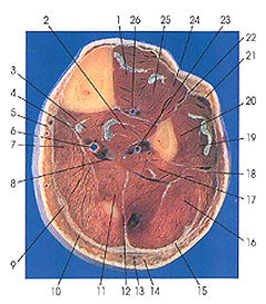
1. Tibialis anterior m. |
8. Tibial nerve |
13. Small saphenous v. |
19. Peroneus longus m. |
This section passes through the anterior, lateral, and posterior compartments of the leg. The gastrocnemius muscle made its last appearance in Plate 7.22, but its aponeurosis is well seen (10, 15). The tendon of plantaris (9) is still located between the aponeuroses of soleus and gastrocnemius. The dominant muscle in the posterior compartment is the soleus muscle (7, 16). The medial and lateral divisions of soleus are divided by a prominent intramuscular septum (12).
The peroneus tertius muscle (23) makes its first appearance in this section. The muscle probably represents, more or less, a completely differentiated, separated portion of extensor digitorum longus. The muscle arises from the distal third of the anterior surface of the fibula, the neighboring interosseous membrane, and the anterior intermuscular septum. The tendon of the muscle inserts onto the base of the fifth metatarsal and often also onto the base of the fourth.
Next Page | Previous Page | Section Top | Title Page
Please send us comments by filling out our Comment Form.
Anatomy Atlases is licensed under a Creative Commons Attribution-NonCommercial-ShareAlike 4.0 International License. 
"Anatomy Atlases", the Anatomy Atlases logo, and "A digital library of anatomy information" are all Trademarks of Michael P. D'Alessandro, M.D.
Anatomy Atlases is funded in whole by Michael P. D'Alessandro, M.D. Advertising is not accepted.
Your personal information remains confidential and is not sold, leased, or given to any third party be they reliable or not.
The information contained in Anatomy Atlases is not a substitute for the medical care and advice of your physician. There may be variations in treatment that your physician may recommend based on individual facts and circumstances.
URL: http://www.anatomyatlases.org/