
Atlas of Human Anatomy in Cross Section: Section 7. Lower Limb
Ronald A. Bergman, Ph.D., Adel K. Afifi, M.D., Jean J. Jew, M.D., and Paul
C. Reimann, B.S.
Peer Review Status: Externally Peer Reviewed
|
Upper Left Quadrant |
Lower Left Quadrant |
Lower Right Quadrant |
Upper Right Quadrant |
|
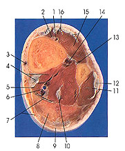
1. Anterior tibial a. and v. and deep peroneal nerve |
5. Flexor digitorum longus m. |
10. Tendon and m. flexor hallucis longus |
13. Interosseous membrane of leg (last appearance) |
This section is three below the preceding one (3 cm). In this section, the bulk of the muscle bellies is decreasing and the tendinous component of each is increasing as the sections approach the ankle region. Note the thickening of the common tendon of the gastrocnemius and soleus muscles (9), which will form the calcaneal (Achilles) tendon. The interosseous membrane (13) is seen for the last time.
The deep peroneal nerve (1) is seen here and for the first time in sections of the lower leg. The deep peroneal nerve arises from the common peroneal. It passes anteriorly and medially through the upper part of the extensor digitorum longus muscle to the interval between that muscle and the tibialis anterior. It descends, in the anterior compartment of the leg, to the ankle, where it divides into terminal branches. In the upper part of the leg, the deep peroneal nerve lies between the extensor digitorum longus and the tibialis anterior muscles and lateral to the anterior tibial artery. In the middle of the leg, it lies anterior to the artery, between extensor hallucis longus and tibialis anterior, and then crosses beneath the extensor hallucis muscle. In the lower third of the leg, It again lies to the lateral side of the artery but between extensor hallucis longus and extensor digitorum longus. It provides muscular branches to tibialis anterior, extensor digitorum longus, extensor hallucis longus, and the peroneus tertius muscles and articular branches to the ankle and lower tibiofibular Joints. The medial terminal branch of the deep peroneal nerve passes distally alongside the dorsalis pedis artery and sends cutaneous branches to the skin between the first and second toes. It also sends articular fibers to the metatarsophalangeal and interphalangeal joints. The lateral terminal branch of the deep peroneal nerve sends fibers to the extensor digitorum brevis muscle and articular twigs to the tarsal joints.
Next Page | Previous Page | Section Top | Title Page
Please send us comments by filling out our Comment Form.
Anatomy Atlases is licensed under a Creative Commons Attribution-NonCommercial-ShareAlike 4.0 International License. 
"Anatomy Atlases", the Anatomy Atlases logo, and "A digital library of anatomy information" are all Trademarks of Michael P. D'Alessandro, M.D.
Anatomy Atlases is funded in whole by Michael P. D'Alessandro, M.D. Advertising is not accepted.
Your personal information remains confidential and is not sold, leased, or given to any third party be they reliable or not.
The information contained in Anatomy Atlases is not a substitute for the medical care and advice of your physician. There may be variations in treatment that your physician may recommend based on individual facts and circumstances.
URL: http://www.anatomyatlases.org/