
Atlas of Human Anatomy in Cross Section: Section 7. Lower Limb
Ronald A. Bergman, Ph.D., Adel K. Afifi, M.D., Jean J. Jew, M.D., and Paul
C. Reimann, B.S.
Peer Review Status: Externally Peer Reviewed
|
Upper Left Quadrant |
Lower Left Quadrant |
Lower Right Quadrant |
Upper Right Quadrant |
|
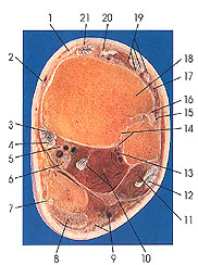
1. Superior extensor retinaculum (transverse crural ligament) |
3. Tendon m. tibialis posterior |
9. Small saphenous v. and sural nerve |
15. Perforating br. of peroneal a. |
This section passes through the lower tibiofibular articulation at about its middle third. The tibiofibular syndesmosis (14) is formed between the distal ends of the tibia and fibula. The two bones are united by the anterior (16) and posterior (14) tibiofibular ligaments and remnants of the interosseous membrane. Occasionally, a synovial articulation occurs in this position. The tendon of the plantaris muscle continues at the medial border of the calcaneal tendon (Achilles) (8). Note that the tendon of extensor digitorum longus has divided into four tendons (19). The lateral compartment muscles (peroneus longus [12] and brevis [11]) now occupy a position posterior to the tibia and fibula, a place shared with flexor hallucis longus (10), the tendon of flexor digitorum longus (4), tibialis posterior (3), the tibial nerve (6), the posterior tibial (5) and lateral calcaneal blood vessels (13), and the calcaneal tendon (Achilles) (8).
Next Page | Previous Page | Section Top | Title Page
Please send us comments by filling out our Comment Form.
Anatomy Atlases is licensed under a Creative Commons Attribution-NonCommercial-ShareAlike 4.0 International License. 
"Anatomy Atlases", the Anatomy Atlases logo, and "A digital library of anatomy information" are all Trademarks of Michael P. D'Alessandro, M.D.
Anatomy Atlases is funded in whole by Michael P. D'Alessandro, M.D. Advertising is not accepted.
Your personal information remains confidential and is not sold, leased, or given to any third party be they reliable or not.
The information contained in Anatomy Atlases is not a substitute for the medical care and advice of your physician. There may be variations in treatment that your physician may recommend based on individual facts and circumstances.
URL: http://www.anatomyatlases.org/