
Atlas of Human Anatomy in Cross Section: Section 7. Lower Limb
Ronald A. Bergman, Ph.D., Adel K. Afifi, M.D., Jean J. Jew, M.D., and Paul
C. Reimann, B.S.
Peer Review Status: Externally Peer Reviewed
|
Upper Left Quadrant |
Lower Left Quadrant |
Lower Right Quadrant |
Upper Right Quadrant |
|
1 . Lateral (third) cuneiform |
5. Calcaneocuboid articulation |
13. Calcaneal tendon (Achilles) |
20. Medial (deltoid) ligament |
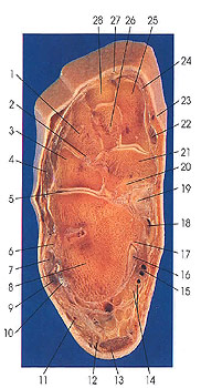
This is the superior (proximal) surface of the next section, looking distally.
This section passes through the calcaneus (8); the navicular (21); the first (25), second (26), and third (1) cuneiform; and the cuboid (3) bones. It cuts the cuneocuboid ligament (2) and the calcaneocuboid articulation (5).
The cuneocuboid ligament (2) connects the apex of the lateral cuneiform with the distal half of the medial surface of the cuboid, Joining proximally with the plantar cuboideonavicular ligament. The calcaneocuboid articulation (5) is the meeting of the reciprocally matched surfaces of the calcaneus and the cuboid bones. The saddle-shaped cuboidal articular surface of the calcaneus meets the posterior articular surface of the cuboid. The articular capsule unites the bones through its attachments to the margins of the articular surfaces, except dorsally, where it is thickened and attached at some distance from the articular margins. Several ligaments are supplementary to the capsule, and these include the calcaneocuboid portion of the bifurcate, the long plantar, and the plantar calcaneocuboid ligaments.
The sustentaculum tali (17) is a well-marked process, located medially on the superior surface of the calcaneus. It projects transversely and bears an elongated concave middle talar articular surface, which matches reciprocally with the middle calcaneal articular surface of the head of the talus. This section passes below the articular surface.
Next Page | Previous Page | Section Top | Title Page
Please send us comments by filling out our Comment Form.
Anatomy Atlases is licensed under a Creative Commons Attribution-NonCommercial-ShareAlike 4.0 International License. 
"Anatomy Atlases", the Anatomy Atlases logo, and "A digital library of anatomy information" are all Trademarks of Michael P. D'Alessandro, M.D.
Anatomy Atlases is funded in whole by Michael P. D'Alessandro, M.D. Advertising is not accepted.
Your personal information remains confidential and is not sold, leased, or given to any third party be they reliable or not.
The information contained in Anatomy Atlases is not a substitute for the medical care and advice of your physician. There may be variations in treatment that your physician may recommend based on individual facts and circumstances.
URL: http://www.anatomyatlases.org/