
Atlas of Human Anatomy in Cross Section: Section 7. Lower Limb
Ronald A. Bergman, Ph.D., Adel K. Afifi, M.D., Jean J. Jew, M.D., and Paul
C. Reimann, B.S.
Peer Review Status: Externally Peer Reviewed
|
Upper Left Quadrant |
Lower Left Quadrant |
Lower Right Quadrant |
Upper Right Quadrant |
|
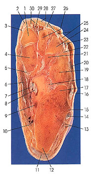
1. First metatarsal |
6. Abductor hallucis m. |
12. Calcaneus |
20. Extensor digitorum brevis m. |
This section passes through the calcaneus (12, 17); first (1), second, third (24), and fourth (22) metatarsals; and the cuboid (19) bones. The peroneal trochlea (15) is seen. Abductor hallucis (6) and quadratus plantae (9) muscles are cut for the first time.
The peroneal (fibular) trochlea of the calcaneus is variable in size and shape and presents a slight groove for the peroneal (fibular) muscles. In this cut, the tendon of peroneus longus (14) is seen behind the trochlea (15).
The abductor hallucis muscle (6) is the largest and most superficial of the intrinsic great toe muscles lying on the medial border of the sole. It passes from the calcaneus across the tendons of the long flexor muscles, runs along the medial side of the short flexor muscle, and is inserted onto the medial side of the base of the proximal phalanx of the great toe and onto the medial side of the long extensor tendon.
The quadratus plantae muscle (9) arises from the medial and plantar surfaces of the calcaneus and is inserted into the lateral margin and deep surface of the tendon of flexor digitorum longus. The muscle has two heads, a small lateral and a larger medial one. There are great individual variations in the structure of this muscle. The lateral head may be missing or greatly reduced in size. The whole muscle may be absent, and the mode of attachment to the flexor digitorum longus tendon varies. It may insert on the flexor tendon of the great toe. There is no homologue in the hand.
Next Page | Previous Page | Section Top | Title Page
Please send us comments by filling out our Comment Form.
Anatomy Atlases is licensed under a Creative Commons Attribution-NonCommercial-ShareAlike 4.0 International License. 
"Anatomy Atlases", the Anatomy Atlases logo, and "A digital library of anatomy information" are all Trademarks of Michael P. D'Alessandro, M.D.
Anatomy Atlases is funded in whole by Michael P. D'Alessandro, M.D. Advertising is not accepted.
Your personal information remains confidential and is not sold, leased, or given to any third party be they reliable or not.
The information contained in Anatomy Atlases is not a substitute for the medical care and advice of your physician. There may be variations in treatment that your physician may recommend based on individual facts and circumstances.
URL: http://www.anatomyatlases.org/