
Atlas of Human Anatomy in Cross Section: Section 7. Lower Limb
Ronald A. Bergman, Ph.D., Adel K. Afifi, M.D., Jean J. Jew, M.D., and Paul
C. Reimann, B.S.
Peer Review Status: Externally Peer Reviewed
|
Upper Left Quadrant |
Lower Left Quadrant |
Lower Right Quadrant |
Upper Right Quadrant |
|
1. Plantar metatarsal aa. |
8. Abductor hallucis m. |
13. Calcaneus, tuberosity |
17. Tendon m. peroneus tertius |
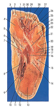
This section passes through the third and fourth layers of the muscles of the foot. It is in the plane of the tendon of peroneus longus (22) and its insertion inferolaterally onto the base and side of the first metatarsal (4). The peroneus longus muscle also inserts onto the inferior surface of the first cuneiform bone. In addition, the muscle usually sends a fibrous slip to the base of the fifth metatarsal. The section cuts the interosseous muscles (25) and other intrinsic foot muscles, including extensor digitorum brevis (2, 26, 29), quadratus plantae (11), and the adductor (16) and abductor hallucis muscles (8). It cuts the tuberosity of the calcaneus (13); the first (4), second (30), third (27), fourth (23), and fifth (20) metatarsals; and the cuboid (19).
Extensor digitorum brevis is the only muscle on the dorsum of the foot. It arises from the dorsal and lateral surfaces of the calcaneus and divides into four muscle bellies whose tendons insert onto the proximal phalanges of the four medial toes just lateral and deep to the tendons of extensor digitorum longus. The belly to the great toe is named extensor hallucis brevis.
Adductor hallucis (16) is composed of two heads, an oblique and a transverse. The oblique head extends from the long plantar ligament to the lateral side of the base of the proximal phalanx of the great toe. Its tendon of insertion is Joined by the transverse head that arises from the capsules of the third, fourth, and fifth metatarsophalangeal joints.
Next Page | Previous Page | Section Top | Title Page
Please send us comments by filling out our Comment Form.
Anatomy Atlases is licensed under a Creative Commons Attribution-NonCommercial-ShareAlike 4.0 International License. 
"Anatomy Atlases", the Anatomy Atlases logo, and "A digital library of anatomy information" are all Trademarks of Michael P. D'Alessandro, M.D.
Anatomy Atlases is funded in whole by Michael P. D'Alessandro, M.D. Advertising is not accepted.
Your personal information remains confidential and is not sold, leased, or given to any third party be they reliable or not.
The information contained in Anatomy Atlases is not a substitute for the medical care and advice of your physician. There may be variations in treatment that your physician may recommend based on individual facts and circumstances.
URL: http://www.anatomyatlases.org/