
Fibula
Ronald A. Bergman, Ph.D., Adel K. Afifi, M.D., Paul M. Heidger,
Jr., Ph.D.
Peer Review Status: Externally Peer Reviewed
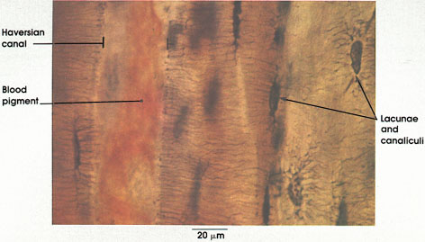
Human, ground bone, unstained, 612 x.
Haversian canal: Conducts blood vessels, lymphatics, and nerves through bone. Haversian canals surrounded by concentric lamellae of compact bone form the Haversian system. These canals are named after Clopton Havers, an English physician, who described them in his Osteologia Novia, published in London in 1691.
Blood pigment: From disintegrated blood elements in the vessels within the Haversian canals.
Lacunae and canalicull: The former are cell spaces that housed osteocytes, and the latter are channels extending out of the lacunae that accommodated cell processes of osteocytes.
Next Page | Previous Page | Section Top | Title Page
Please send us comments by filling out our Comment Form.
All contents copyright © 1995-2024 the Author(s) and Michael P. D'Alessandro, M.D. All rights reserved.
"Anatomy Atlases", the Anatomy Atlases logo, and "A digital library of anatomy information" are all Trademarks of Michael P. D'Alessandro, M.D.
Anatomy Atlases is funded in whole by Michael P. D'Alessandro, M.D. Advertising is not accepted.
Your personal information remains confidential and is not sold, leased, or given to any third party be they reliable or not.
The information contained in Anatomy Atlases is not a substitute for the medical care and advice of your physician. There may be variations in treatment that your physician may recommend based on individual facts and circumstances.
URL: http://www.anatomyatlases.org/