
Plate 17.318 Spinal Cord
Ronald A. Bergman, Ph.D., Adel K. Afifi, M.D., Paul M. Heidger,
Jr., Ph.D.
Peer Review Status: Externally Peer Reviewed
Human, Müller's fluid, Weigert's method, 10 x.
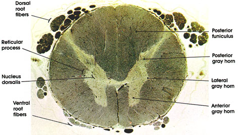
Dorsal root fibers: Central processes of dorsal root ganglia.
Reticular process: Characteristic of cervical levels of the spinal cord but also located at other spinal levels. Located between the posterior and anterior horns and produced by an extension of gray matter into the adjacent white substance. Constitutes the lateral zone of lamina V of Rexed. See also Plate 317.
Nucleus dorsallis: Distinct nuclear mass located in the medial part of the base of the posterior horn. In this nucleus, dorsal root fibers synapse with neurons destined to form the dorsal (posterior) spinocerebellar tract. The nucleus extends between C8 and L2 spinal segments. Also known as the column of Clarke*.
Ventral root fibers: See Plates 114, 322, and 323.
Posterior funiculus: The white matter of the cord located between the posterior central (median) septum and the medial border of the posterior horn. Contains heavily myelinated fibers that form the gracile and cuneate tracts. Note the large size of this funiculus at this level compared to lower levels of the spinal cord. See also Plates 316, 317, and 319.
Posterior gray horn: A mass of neurons in the posterolateral part of the spinal cord. Receive collaterals or terminals of dorsal root fibers. Sends axons to anterior horn cells, interneurons, or to ascending tracts. See also Plates 316 and 323.
Lateral gray horn: Characteristic of thoracic level, this projection is formed by the intermediolateral nucleus. Contains visceral efferent neurons of the sympathetic nervous system. Extends from C8 to L2-4. Axons of neurons here exit with the ventral horn fibers to terminate in the chain of ganglia (sympathetic), where they synapse with ganglion cells whose axons are widely distributed to the iris (dilator smooth muscle); lacrimal, salivary and sweat glands; bronchi; heart; smooth muscle of the gastrointestinal tract; sex organs; urinary bladder; adrenal medulla; and blood vessels.
Anterior gray horn: A mass of large neurons in the anterolateral part of the spinal cord. Contains somatic efferent neurons. Compare the size of the horn at this level with those seen at higher and lower levels. See also Plates 316, 317, and 323.
*Clarke, 1817-1880, was an English anatomist and physician.
Next Page | Previous Page | Section Top | Title Page
Please send us comments by filling out our Comment Form.
All contents copyright © 1995-2024 the Author(s) and Michael P. D'Alessandro, M.D. All rights reserved.
"Anatomy Atlases", the Anatomy Atlases logo, and "A digital library of anatomy information" are all Trademarks of Michael P. D'Alessandro, M.D.
Anatomy Atlases is funded in whole by Michael P. D'Alessandro, M.D. Advertising is not accepted.
Your personal information remains confidential and is not sold, leased, or given to any third party be they reliable or not.
The information contained in Anatomy Atlases is not a substitute for the medical care and advice of your physician. There may be variations in treatment that your physician may recommend based on individual facts and circumstances.
URL: http://www.anatomyatlases.org/