
Plate 17.338 Mesencephalon
Ronald A. Bergman, Ph.D., Adel K. Afifi, M.D., Paul M. Heidger,
Jr., Ph.D.
Peer Review Status: Externally Peer Reviewed
Human, 10% formalin, Pal-Weigert, 2.7 x.
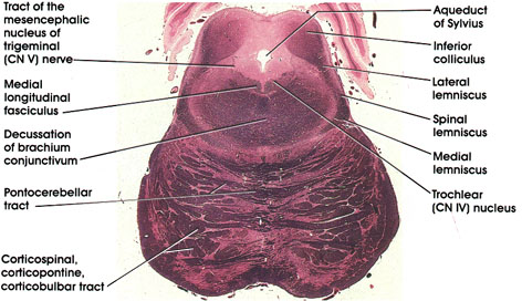
Aqueduct of Sylvius: Connecting the third and fourth ventricles. Sylvius (Jacques Dubois) was a sixteenth -century French anatomist.
Trochlear (CN IV) nucleus: Motor neurons located in a paramedian position dorsal to the medial longitudinal fasciculus. Axons of neurons in trochlear nucleus decussate prior to leaving the neuraxis (see Plate 337).
Inferior colliculus: Ovoid cellular mass in the tecturn of the mesencephalon. Belongs to the auditory system.
Lateral lemniscus: Located laterally and dorsally as it enters the inferior colliculus. Concerned with audition.
Spinal and medial lemnisci: Continuation of the same structures seen at more caudal levels.
Corticospinal, corticopontine, corticobulbar tracts: Sectioned transversely on their way to pontine nuclei, cranial nerve nuclei, and motor neurons of the spinal cord.
Pontocerebellar tract: Axons of pontine nuclei on their way to the cerebellum.
Decussation of brachium conjunctivum: Massive outflow tract of the cerebellum seen decussating at this level. Fibers project, after decussation, into the red nucleus and ventral lateral nucleus of the thalamus.
Tract of the mesencephalic nucleus of trigerninal (CN V) nerve: Processes of pseudounipolar neurons in the mesencephalic nucleus of the trigeminal nerve. Neurons are sparsely scattered on each side of the tract.
Medial longitudinal fasciculus: Continuation of the same structure seen at more rostral and more caudal levels.
Next Page | Previous Page | Section Top | Title Page
Please send us comments by filling out our Comment Form.
All contents copyright © 1995-2024 the Author(s) and Michael P. D'Alessandro, M.D. All rights reserved.
"Anatomy Atlases", the Anatomy Atlases logo, and "A digital library of anatomy information" are all Trademarks of Michael P. D'Alessandro, M.D.
Anatomy Atlases is funded in whole by Michael P. D'Alessandro, M.D. Advertising is not accepted.
Your personal information remains confidential and is not sold, leased, or given to any third party be they reliable or not.
The information contained in Anatomy Atlases is not a substitute for the medical care and advice of your physician. There may be variations in treatment that your physician may recommend based on individual facts and circumstances.
URL: http://www.anatomyatlases.org/