
Plate 17.341 Mesencephalon-Diencephalon Junction
Ronald A. Bergman, Ph.D., Adel K. Afifi, M.D., Paul M. Heidger,
Jr., Ph.D.
Peer Review Status: Externally Peer Reviewed
Human, 10% formalin, Pal-Welgert, 2.3 x.
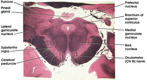
Pulvinar: Belongs to the lateral group of thalamic nuclei. Has reciprocal connections with the medial and lateral geniculate bodies caudally and the association parietal, temporal, and occipital cortices rostrally. Plays a role in several neural functions, including vision, audition, speech, and pain.
Lateral geniculate body: A thalamic relay nucleus concerned with vision. Receives fibers from the optic tract and projects to the primary visual cortex.
Substantia nigra: A mass of pigmented cells containing melanin located dorsal to the cerebral peduncle. This area is invariably the site of pathologic changes associated with Parkinson's disease.
Cerebral peduncle: Descending corticofugal fiber system. Lesion results in contralateral muscle weakness or paralysis.
Oculomotor (CN III) nerve: Coursing in the tegmentum of the midbrain medial to the substantia nigra and cerebral peduncle.
Red nucleus: So-called because of a pinkish color in the fresh state owing to its high vascularity. Links the cerebellum, cerebral cortex, and spinal cord.
Medial geniculate body: A thalamic relay nucleus concerned with audition. Receives fibers from brachium of the inferior colliculus and projects to the primary auditory cortex.
Brachium of superior colliculus: Fiber bundle connecting the superior colliculus and the lateral geniculate nucleus.
Pretectal nucleus: Rostral extension of the superior colliculus. Receives optic tract fibers and projects bilaterally to oculornotor nuclei. Important relay in pupillary light reflex.
Pineal gland: Located dorsal to the mesencephalon. Part of the epithalamus. Has endocrine function and is an important landmark radiologically.
Next Page | Previous Page | Section Top | Title Page
Please send us comments by filling out our Comment Form.
All contents copyright © 1995-2024 the Author(s) and Michael P. D'Alessandro, M.D. All rights reserved.
"Anatomy Atlases", the Anatomy Atlases logo, and "A digital library of anatomy information" are all Trademarks of Michael P. D'Alessandro, M.D.
Anatomy Atlases is funded in whole by Michael P. D'Alessandro, M.D. Advertising is not accepted.
Your personal information remains confidential and is not sold, leased, or given to any third party be they reliable or not.
The information contained in Anatomy Atlases is not a substitute for the medical care and advice of your physician. There may be variations in treatment that your physician may recommend based on individual facts and circumstances.
URL: http://www.anatomyatlases.org/