
Plate 17.355 Anterior Commissure
Ronald A. Bergman, Ph.D., Adel K. Afifi, M.D., Paul M. Heidger,
Jr., Ph.D.
Peer Review Status: Externally Peer Reviewed
Human, 10% formalin, Weigert's hematoxylin (Loyez), 1.1 x.
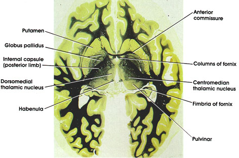
Columns of fornix: Body of fornix separates into two columns, each of which curves ventrally and courses through the hypothalamus on its way to the mamillary body.
Anterior commissure: A compact fiber bundle in close proximity to anterior extent of the fornix. Interconnects olfactory bulbs and the temporal cortices.
Centromedion thalamic nucleus: Belongs to the intralaminar group of thalamic nuclei. Receives fibers from several sources, motor and sensory, that project diffusely to cerebral cortex either directly or indirectly via other thalamic nuclei. Plays a role in cortical arousal response and in pain mechanism.
Pulvinar: Belongs to the lateral group of thalamic nuclei. Has reciprocal connections with the medial and lateral geniculate bodies caudally and the association parietal, temporal, and occipital cortices rostrally. Plays a role in several neural functions, including vision, audition, speech, and pain.
Fimbria of fornix: Efferent fibers from the hippocampus that merge with the fornix.
Putamen: Lateral to globus pallidus. One of the basal ganglia nuclei. Concerned with motor control.
Globus pallidus: Medial to the putamen. Separated from the thalamus by the posterior limb of the internal capsule. Has two segments: outer, close to the putamen, and inner, close to the internal capsule. Characterized by heavily myelinated fibers traversing it. A component of the basal ganglia. Concerned with motor control.
Internal capsule (posterior limb): Separates putamen and globus pallidus from thalamus. Carries motor and sensory fibers from and to the cerebral cortex.
Dorsomedial thalamic nucleus: Belongs to the medial group of thalamic nuclei. Most highly developed in man. Concerned with affective behavior and memory.
Habenula: A nuclear mass located dorsal to the thalamus at the junction of the diencephalon and midbrain. A component of the limbic system.
Next Page | Previous Page | Section Top | Title Page
Please send us comments by filling out our Comment Form.
All contents copyright © 1995-2024 the Author(s) and Michael P. D'Alessandro, M.D. All rights reserved.
"Anatomy Atlases", the Anatomy Atlases logo, and "A digital library of anatomy information" are all Trademarks of Michael P. D'Alessandro, M.D.
Anatomy Atlases is funded in whole by Michael P. D'Alessandro, M.D. Advertising is not accepted.
Your personal information remains confidential and is not sold, leased, or given to any third party be they reliable or not.
The information contained in Anatomy Atlases is not a substitute for the medical care and advice of your physician. There may be variations in treatment that your physician may recommend based on individual facts and circumstances.
URL: http://www.anatomyatlases.org/