
Atlas of Human Anatomy in Cross Section: Section 3. Middle Thorax (Heart and Lungs)
Ronald A. Bergman, Ph.D., Adel K. Afifi, M.D., Jean J. Jew, M.D., and Paul
C. Reimann, B.S.
Peer Review Status: Externally Peer Reviewed
|
Upper Left Quadrant |
Lower Left Quadrant |
Lower Right Quadrant |
Upper Right Quadrant |
|
1. Rib 7 and anterior mediastinal fat and cardiac fat on right atrium |
13. Liver, right lobe |
26. Supraspinous ligament |
39. Central tendon of diaphragm |
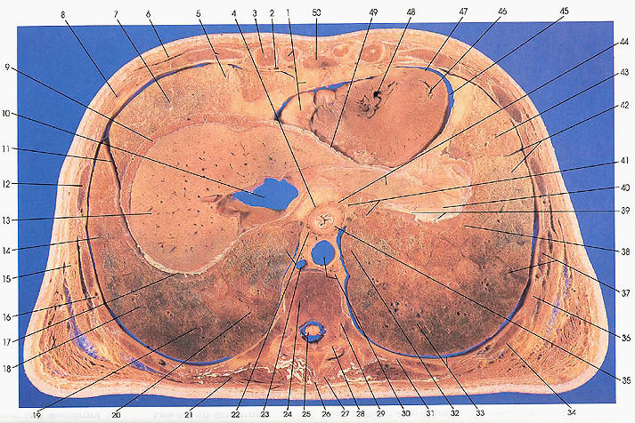
This section passes through the tenth thoracic vertebra (24), its costovertebral articulation (29), and the intervertebral articulation, T9/10 (28). It cuts ribs 10 (31), 9 (34), 8 (37), 7 (12), 6 (8), and 5 (5) and the sixth (3) and seventh (1) costal cartilages. Note that the costocartilage does not articulate with the sternum (xiphoid process) at this level. The section passes through the right and left domes of the diaphragm, penetrating more deeply on the right side. The cut edges of the diaphragm (9, 41), and portions of the right (13) and left (40) lobes of the liver can be seen. The left lobe of the liver, in this subject, is a large persistent remnant of development that terminates near the spleen as a fibrous band (appendix fibrosa hepatic). This is called a "beaver tailed" liver.
The middle mediastinum contains the heart, including the right ventricle (48), apex (47), left ventricle (46), pericardial cavity (45), and middle mediastinal fat (1).
On the left side, the following left lung bronchopulmonary segments can be seen: left upper lobe, lower division (lingular), inferior (43); left lower lobe, anterior medial basal (42); left lower lobe, lower division (lingular), inferior (41); left upper lobe, lower division, superior segment (38); left lower lobe, lateral basal (37), left lower lobe, posterior basal (33); and left lower lobe, anterior medial basal (32).
The posterior mediastinum contains the esophagus (35) anteriorly and the thoracic aorta (30) posteriorly, flanked by the azygos vein (23) on the right and small lymphatic and blood vessels and elements of the autonomic nervous system (30) on the left.
On the right side, the bronchopulmonary segments of the right lung include the right middle lobe, medial (22); right lower lobe, medial basal (20); right lower lobe, posterior basal (19); right lower lobe, lateral basal (18); right lower lobe, anterior basal (14); right middle lobe, lateral (7); and right middle lobe, medial (5).
The thorax, middle portion, ends with this cut.
Next Page | Previous Page | Section Top | Title Page
Please send us comments by filling out our Comment Form.
Anatomy Atlases is licensed under a Creative Commons Attribution-NonCommercial-ShareAlike 4.0 International License. 
"Anatomy Atlases", the Anatomy Atlases logo, and "A digital library of anatomy information" are all Trademarks of Michael P. D'Alessandro, M.D.
Anatomy Atlases is funded in whole by Michael P. D'Alessandro, M.D. Advertising is not accepted.
Your personal information remains confidential and is not sold, leased, or given to any third party be they reliable or not.
The information contained in Anatomy Atlases is not a substitute for the medical care and advice of your physician. There may be variations in treatment that your physician may recommend based on individual facts and circumstances.
URL: http://www.anatomyatlases.org/