
Atlas of Human Anatomy in Cross Section: Section 4. Upper Limb
Ronald A. Bergman, Ph.D., Adel K. Afifi, M.D., Jean J. Jew, M.D., and Paul
C. Reimann, B.S.
Peer Review Status: Externally Peer Reviewed
|
Upper Left Quadrant |
Lower Left Quadrant |
Lower Right Quadrant |
Upper Right Quadrant |
|
1. Cephalic v. |
4. Radial nerve |
9. Triceps brachii, lateral head m. |
14. Basilic v. and anastomotic branch |
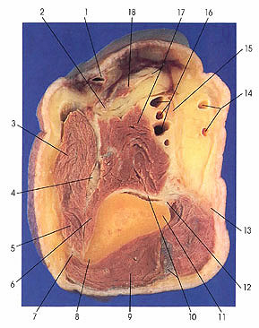
This section passes through the medial (11) and lateral (8) epicondyles of the humerus. The medial (12) and lateral (7) intermuscular septa are seen separating the anterior and posterior compartments.
Note the position of brachioradialis (3) and extensor carpi radialis longus (5) muscles anterior to the lateral epicondyle from which they both arise. Because of their position above the elbow joint, these two muscles are functionally flexors but are classified as extensors, in part because they are innervated by the radial nerve (4). Extensor carpi radialis longus muscle (5) is seen for the first time in this section.
Note that the ulnar nerve (13) has almost completed its move dorsal to the humerus where it will lie in the ulnar sulcus, a groove on the dorsal surface of the medial epicondyle.
The tendon of biceps brachii (18) is composed of two parts: the bicipital aponeurosis or lacertus fibrosus (17), which extends medially to join the antebrachial fascia on the ulnar side of the forearm, and a tough band of tendon (2) that descends into the space between brachioradialis (3) and pronator teres (seen in next section) muscles and above the receding brachialis muscle (17), which inserts onto the dorsal half of the bicipital tuberosity of the radius.
Next Page | Previous Page | Section Top | Title Page
Please send us comments by filling out our Comment Form.
Anatomy Atlases is licensed under a Creative Commons Attribution-NonCommercial-ShareAlike 4.0 International License. 
"Anatomy Atlases", the Anatomy Atlases logo, and "A digital library of anatomy information" are all Trademarks of Michael P. D'Alessandro, M.D.
Anatomy Atlases is funded in whole by Michael P. D'Alessandro, M.D. Advertising is not accepted.
Your personal information remains confidential and is not sold, leased, or given to any third party be they reliable or not.
The information contained in Anatomy Atlases is not a substitute for the medical care and advice of your physician. There may be variations in treatment that your physician may recommend based on individual facts and circumstances.
URL: http://www.anatomyatlases.org/