
Atlas of Human Anatomy in Cross Section: Section 4. Upper Limb
Ronald A. Bergman, Ph.D., Adel K. Afifi, M.D., Jean J. Jew, M.D., and Paul
C. Reimann, B.S.
Peer Review Status: Externally Peer Reviewed
|
Upper Left Quadrant |
Lower Left Quadrant |
Lower Right Quadrant |
Upper Right Quadrant |
|
1. Tendon m. palmaris longus |
8. Brachioradialis m. |
14. Anterior interosseous a. and v. |
23. Basilic v. |
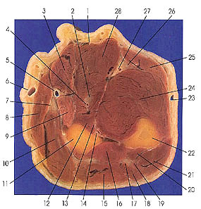
This section passes through the upper middle third of the forearm.
The radial artery (5) is located between brachioradialis (8) and flexor carpi radialis (3) muscles. It is also lateral to flexor carpi radialis in the distal forearm, hence the muscle is a guide to finding the radial artery.
The muscles of the ventral forearm are found in four layers. The first layer is composed of four muscles that arise partly from the medial epicondyle of the humerus and partly from the ulna. Pronator teres (9) is a band like, strong muscle that inserts onto the middle third of the shaft of the radius. Flexor carpi radialis (3) is a fusiform muscle that inserts onto the base of the second metacarpal bone. Palmaris longus (1), when present, is inserted into the palmer aponeurosis. Flexor carpi ulnaris (25) is inserted onto the pisiform bone and palmer fascia. Flexor carpi ulnaris is innervated by the ulnar nerve (26), and the other three are supplied by the median nerve (2).
Flexor digitorum superficialis (28) is considered to be the only muscle of the second layer of muscles on the ventral side of the forearm. This muscle is deep to the first layer but becomes superficial in the distal forearm. It arises from the medial epicondyle of the humerus and from the ulna (22) and radius (11). Its tendons split opposite the middle phalanx of each finger to allow the tendons of flexor digitorum profundus (24) to pass to their attachments on the distal phalanges. It is supplied by the median nerve (2).
The third layer is composed of two muscles, but it is believed that they differentiate from a single deep flexor muscle. Flexor digitorum profundus (24) arises from the upper three fourths of the ventral and medial surfaces of the ulna. Its four tendons enter osteofibrous tunnels of the fingers, pass through slits in superficialis tendons, and insert onto the bases of the distal phalanges. Flexor pollicis longus (12) is broad and flat, arises from the ventral surface of the radius, and inserts onto the base of the distal phalanx of the thumb. Both muscles are innervated by the median nerve (2), but the ulnar half of flexor profundus (24) is also supplied by the ulnar nerve (26).
The fourth layer is composed of a single muscle, pronator quadratus, which is not seen at this level. This muscle passes transversely across the lower forearm from ulna to radius. It is innervated by the anterior interosseous branch (13) of the median nerve.
Next Page | Previous Page | Section Top | Title Page
Please send us comments by filling out our Comment Form.
Anatomy Atlases is licensed under a Creative Commons Attribution-NonCommercial-ShareAlike 4.0 International License. 
"Anatomy Atlases", the Anatomy Atlases logo, and "A digital library of anatomy information" are all Trademarks of Michael P. D'Alessandro, M.D.
Anatomy Atlases is funded in whole by Michael P. D'Alessandro, M.D. Advertising is not accepted.
Your personal information remains confidential and is not sold, leased, or given to any third party be they reliable or not.
The information contained in Anatomy Atlases is not a substitute for the medical care and advice of your physician. There may be variations in treatment that your physician may recommend based on individual facts and circumstances.
URL: http://www.anatomyatlases.org/