
Atlas of Human Anatomy in Cross Section: Section 7. Lower Limb
Ronald A. Bergman, Ph.D., Adel K. Afifi, M.D., Jean J. Jew, M.D., and Paul
C. Reimann, B.S.
Peer Review Status: Externally Peer Reviewed
|
Upper Left Quadrant |
Lower Left Quadrant |
Lower Right Quadrant |
Upper Right Quadrant |
|
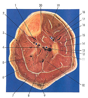
1. Posterior tibial vessels |
4. Tibial nerve |
9. Gastrocnemius muscle (lateral head) and small saphenous v. |
15. Superficial peroneal nerve |
This section is three below the preceding one (3 cm).
The popliteal artery has divided into two branches that supply different parts of the leg. The anterior tibial artery (17) supplies the anterior compartment muscles, and the posterior tibial artery (1) supplies the posterior compartment muscles. The posterior tibial artery gives rise to the peroneal artery (12), which supplies structures on the posterolateral side of the leg.
The tendon of plantaris (5) is now a flattened band and occupies an interval between the deep fascia of gastrocnemius (7) and soleus (10) muscles.
The sural nerve (8), at this level, lies behind the gastrocnemius muscle at the fascial plane marking the division between the lateral (9) and medial (7) heads of the gastrocnemius muscle.
The deep peroneal nerve (17) arises from the common peroneal and supplies muscular branches to tibialis anterior (19), extensor digitorum longus (18), extensor hallucis longus (18), and peroneus tertius muscles. The superficial peroneal nerve (15) gives muscular branches to peroneus longus and peroneus brevis muscles. The remainder of the nerve is sensory to the skin of the lower aspect of the leg and the dorsum of the foot.
The anterior (16) and posterior (11) crural septa and the fibula define the lateral compartment and its muscular content.
The lateral compartment muscles consist of two muscles: peroneus brevis (14) and peroneus longus (13). Peroneus brevis arises from the middle third of the lateral surface of the fibula and from the septa (11, 16) that separate it from the anterior and posterior groups of muscles. The muscle inserts onto the dorsal aspect of the tuberosity of the fifth metatarsal. The anterior head (tendinous) of peroneus longus arises from the anterior capitular ligament (see Plate 7.17, 18), the neighboring part of the lateral condyle of the tibia, and the head of the fibula (see Plate 7.17, 16); the fleshy anterior head, from the proximal third of the anterior intermuscular septum (16) and the crural fascia; and the posterior fleshy head, from the proximal half of the lateral surface of the shaft of the fibula and the posterior intermuscular septum (11). The muscle inserts on the inferior surface of the first cuneiform and on the adjacent part of the inferolateral border and base of the first metatarsal. A fibrous slip is usually sent to the base of the fifth metatarsal. This fibrous slip arises from the tendon of peroneus longus within the groove of the cuboid bone.
Next Page | Previous Page | Section Top | Title Page
Please send us comments by filling out our Comment Form.
Anatomy Atlases is licensed under a Creative Commons Attribution-NonCommercial-ShareAlike 4.0 International License. 
"Anatomy Atlases", the Anatomy Atlases logo, and "A digital library of anatomy information" are all Trademarks of Michael P. D'Alessandro, M.D.
Anatomy Atlases is funded in whole by Michael P. D'Alessandro, M.D. Advertising is not accepted.
Your personal information remains confidential and is not sold, leased, or given to any third party be they reliable or not.
The information contained in Anatomy Atlases is not a substitute for the medical care and advice of your physician. There may be variations in treatment that your physician may recommend based on individual facts and circumstances.
URL: http://www.anatomyatlases.org/