
Atlas of Human Anatomy in Cross Section: Section 7. Lower Limb
Ronald A. Bergman, Ph.D., Adel K. Afifi, M.D., Jean J. Jew, M.D., and Paul
C. Reimann, B.S.
Peer Review Status: Externally Peer Reviewed
|
Upper Left Quadrant |
Lower Left Quadrant |
Lower Right Quadrant |
Upper Right Quadrant |
|
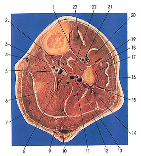
1 . Interosseous membrane of leg |
6. Tendon m. plantaris |
11. Soleus m. |
17. Peroneus brevis m. |
This section is three below the preceding one (3 cm).
All of the musculature of the back of the leg, except the popliteus muscle, is represented in this section. Those of the superficial group include gastrocnemius (7, 12), soleus (11), and plantaris, represented by its long thin tendon (6). Those of the deep group include flexor digitorum longus (2) and flexor hallucis longus (14). Flexor hallucis longus makes its first appearance in this section. Popliteus muscle also belongs to the deep group, and it may be seen in Plate 7.17 (8) and Plate 7.18 (2).
The medial head (the broader and thicker of the two heads) of gastrocnemius (7) arises from the back of the medial condyle of the femur above the articular surface, from an area on the back of the femur superolateral to the condylar origin, and from the femoral margin of the capsule of the knee joint. The lateral head (12) arises from a facet on the proximal portion of the posterolateral surface of the lateral condyle of the femur and from a proximomedial area above the lateral condyle. The insertion of gastrocnemius is discussed following the description of the origin of the soleus muscle.
The fibular head of soleus (11) arises from the back of the head of the fibula (see Plate 7.17, 14) and from the proximal third of the posterior surface of the shaft of the fibula, from the intermuscular septum (15) between it and the peroneus longus (16). The tibial head arises from the popliteal line and from the middle third of the medial border of the tibia.
The calcaneal tendon (Achilles) is the common tendon of gastrocnemius and soleus (triceps surae), and it is the thickest, hence the strongest, in the body. The tendon begins as a broad aponeurosis on the deep side of each muscle (3, 8) and passes with muscle bundles obliquely, from each side, to insert in a bipenniform manner on the deep surface of the calcaneal tendon. From the distal half of the leg the cancaneal tendon begins as a broad aponeurosis that covers the greater part of the posterior surface of the gastrocnemius muscle, which gradually converges into the heavy fibrous band (tendon) that is inserted onto the calcaneus bone of the foot.
Next Page | Previous Page | Section Top | Title Page
Please send us comments by filling out our Comment Form.
Anatomy Atlases is licensed under a Creative Commons Attribution-NonCommercial-ShareAlike 4.0 International License. 
"Anatomy Atlases", the Anatomy Atlases logo, and "A digital library of anatomy information" are all Trademarks of Michael P. D'Alessandro, M.D.
Anatomy Atlases is funded in whole by Michael P. D'Alessandro, M.D. Advertising is not accepted.
Your personal information remains confidential and is not sold, leased, or given to any third party be they reliable or not.
The information contained in Anatomy Atlases is not a substitute for the medical care and advice of your physician. There may be variations in treatment that your physician may recommend based on individual facts and circumstances.
URL: http://www.anatomyatlases.org/