
Atlas of Human Anatomy in Cross Section: Section 7. Lower Limb
Ronald A. Bergman, Ph.D., Adel K. Afifi, M.D., Jean J. Jew, M.D., and Paul
C. Reimann, B.S.
Peer Review Status: Externally Peer Reviewed
|
Upper Left Quadrant |
Lower Left Quadrant |
Lower Right Quadrant |
Upper Right Quadrant |
|
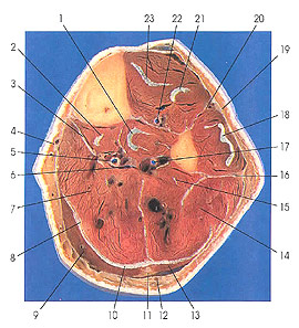
1. Tendon and m. tibialis posterior |
6. Tibial nerve |
12. Small saphenous v. |
17. Peroneal a. |
This section passes through the middle of the leg, three sections below the preceding one (3 cm).
The posterior compartment of the leg is now largely composed of the soleus muscle (7, 14), although gastrocnemius (lateral [13] and medial [9] heads), flexor hallucis longus (15), flexor digitorum longus (2), and tibialis posterior (1) muscles are seen. The greatest reduction in the fleshy bulk of the leg is due to the reduction in size of the gastrocnemius muscle. Gastrocnemius makes its last appearance in this section.
The deep muscles of the posterior compartment of the leg include the popliteus muscle, which was discussed with Plate 7.18. Flexor digitorum longus (2) arises from the popliteal line, the medial side of the second quarter of the dorsal surface of the tibia, the fibrous septum between the muscle and tibialis posterior, and the fascia covering its proximal extremity. The tendon of insertion passes behind the medial malleolus, dorsolateral to the tendon of tibialis posterior, crosses the posterior talotibial ligament, and passes along the medial margin of the sustentaculum tali into the sole of the foot. Here it crosses the tendon of flexor hallucis longus, from which it receives a tendinous slip. It divides into four parts that pass to the second, third, fourth, and fifth toes. Each tendon is held in place on the phalanges of the toe to which it passes by a fibrous sheath. Superficial to it, in the sheath, lies a tendon of flexor digitorum brevis, which bifurcates for the tendon of flexor digitorum longus as it passes to the base of the terminal phalanx. The tendon is connected, like the corresponding tendons of the fingers, by vincula tendinum, to the phalanges of the toes.
Flexor hallucis longus (15) arises from the distal two-thirds of the posterior surface of the fibula and from the septa between it and tibialis posterior and the peroneal muscles. The tendon of flexor hallucis longus passes behind the ankle joint and enters the groove on the posterior surface of the talus and the undersurface of the sustentaculum tall, where it lies on the fibular side of the tendon of flexor digitorum longus. It then crosses the deep surface of this tendon, to which it gives a slip, passes onto the plantar surface of the medial head of flexor hallucis brevis, and between the sesamoid bones of this muscle into the osseofibrous tunnel to insert on the plantar surface of the big toe.
Tibialis posterior (1) arises from the lateral half of the popliteal line and the lateral half of the middle third of the posterior surface of the tibia; from the medial side of the head and of that part of the fibula adjacent to the interosseous membrane in the proximal two-thirds of the leg; from the whole of the proximal and lateral portion of the distal part of the posterior surface of the interosseous membrane; and from the septa between its proximal portion and the long flexor muscles. The tendon of insertion divides into two divisions, deep and superficial. The deep portion becomes attached chiefly to the tubercle of the navicular bone, and usually to the first cuneiform. The superficial tendon spreads out to be attached chiefly to the third cuneiform and the base of the fourth metatarsal, but also in part to the second cuneiform, to the capsule of the naviculocuneiform joint, to the sulcus of the cuboid, and usually also to the origin of the short flexor of the big toe and the base of the second metatarsal.
Next Page | Previous Page | Section Top | Title Page
Please send us comments by filling out our Comment Form.
Anatomy Atlases is licensed under a Creative Commons Attribution-NonCommercial-ShareAlike 4.0 International License. 
"Anatomy Atlases", the Anatomy Atlases logo, and "A digital library of anatomy information" are all Trademarks of Michael P. D'Alessandro, M.D.
Anatomy Atlases is funded in whole by Michael P. D'Alessandro, M.D. Advertising is not accepted.
Your personal information remains confidential and is not sold, leased, or given to any third party be they reliable or not.
The information contained in Anatomy Atlases is not a substitute for the medical care and advice of your physician. There may be variations in treatment that your physician may recommend based on individual facts and circumstances.
URL: http://www.anatomyatlases.org/