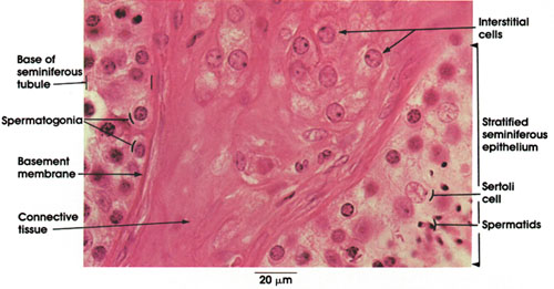

Plate 14.268 Interstitial Cells
Ronald A. Bergman, Ph.D., Adel K. Afifi, M.D., Paul M. Heidger,
Jr., Ph.D.
Peer Review Status: Externally Peer Reviewed

Human, 10% formalin, H. & E., 612 x.
This figure shows parts of two seminiferous tubules separated by a connective tissue sheath. Within this connective tissue sheath are embedded large ovoid cells, the interstitial cells of Leydig. These occur in groups, have a rounded, large eccentric nucleus with a prominent nucleolus and a vacuolated cytoplasm, which results from the loss of lipid droplets and crystals during tissue processing. The interstitial cells are the source of testosterone, the male sex hormone, whose functions include the development and maintenance of secondary sex characteristics and the structure and function of the male accessory organs, the development of psychosexual behavior (in part) in the mature male, a role in protein metabolism, and the regulation of the output of the pituitary gonadotropic hormone, interstitial cell-stimulating hormone (ICSH). The seminiferous tubules shown reveal part of their contents, spermatogonia, spermatids, and Sertoli cells (see Plates 21, 264, and 265).
Next Page | Previous Page | Section Top | Title Page
Please send us comments by filling out our Comment Form.
All contents copyright © 1995-2025 the Author(s) and Michael P. D'Alessandro, M.D. All rights reserved.
"Anatomy Atlases", the Anatomy Atlases logo, and "A digital library of anatomy information" are all Trademarks of Michael P. D'Alessandro, M.D.
Anatomy Atlases is funded in whole by Michael P. D'Alessandro, M.D. Advertising is not accepted.
Your personal information remains confidential and is not sold, leased, or given to any third party be they reliable or not.
The information contained in Anatomy Atlases is not a substitute for the medical care and advice of your physician. There may be variations in treatment that your physician may recommend based on individual facts and circumstances.
URL: http://www.anatomyatlases.org/