
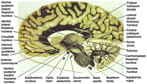
Plate 17.363 Section through Centromedian Thalamic Nucleus
Ronald A. Bergman, Ph.D., Adel K. Afifi, M.D., Paul M. Heidger,
Jr., Ph.D.
Peer Review Status: Externally Peer Reviewed
Human, 10% formalin, Weigert's hematoxylin (Loyez), 1 x.
Internal capsule (posterior limb): The part of the internal capsule separating the thalamus from the basal ganglia. Contains fibers destined for and leaving the cerebral cortex.
Caudatenucleus: One of the basal ganglia nuclei. Concerned with motor control. Note the characteristic bulge into the cavity of the lateral ventricle.
Anterior commissure: A compact fiber bundle connecting the olfactory bulbs and the temporal cortices.
Ventral posterior medial thalamic nucleus: One of the lateral group of thalamic nuclei. Receives the trigeminothalamic fiber system, including taste fibers, and projects to the primary somesthetic cortex in the postcentral gyrus.
Optic tract: Conveys impulses from the contralateral visual field to the lateral geniculate nucleus and the pretectal area.
Cerebral peduncle: A bundle of corticofugal fibers located inferior to the substantia nigra in the midbrain. Contains corticospinal, corticobulbar, and corticopontine fiber bundles. Lesions produce weakness or paralysis in the contralateral half of the body, including the face.
Oculomotor nerve: Seen leaving the ventral surface of the midbrain.
Subthalamic nucleus: Also known as corpus Luysii. Shaped like a biconcave lens. Receives fibers from and projects to the globus pallidus. Discrete lesions here result in an abnormal type of flinging movement known as ballism.
Basis pontis: Contains descending corticofugal fibers, pontine nuclei, and pontocerebellar fibers.
Restiform body: Also known as inferior cerebellar peduncle. A compact bundle of nerve fibers connecting the medulla with the cerebellum. Tracts and fibers forming this bundle originate in the medulla and the spinal cord.
Dentate nucleus: One of the deep cerebellar nuclei. Receives fibers from the Purkinje neurons of the cerebellar hemispheres and projects via the dentatorubrothalarnic system to the contralateral red nucleus and ventrolateral nucleus of the thalamus. Concerned with motor control.
Substantia nigra: The largest nuclear mass in the midbrain. Contains pigmented neurons (melanin). Connected with basal ganglia and thalamus. important in motor control. This area is invariably the site of pathological changes associated with Parkinson's disease.
Centromedian (thalamic) nucleus: Belongs to the intralaminar group of thalamic nuclei. Concerned with a variety of sensory and motor functions and arousal.
Pulvinar: One of the lateral group of thalamic nuclei. Has reciprocal connections with the medial and lateral geniculate bodies caudally and association parietal, temporal, and occipital cortices rostrally. involved in several neural functions, including vision, audition, speech, and pain.
Corpus callosum (splenium): The posterior part of the corpus callosurn. Concerned with transfer of visual information between the hemispheres.
Fornix (crus): Note the relationship of the crus of the fornix to the splenium of the corpus callosurn. The crus is a continuation of the fimbria of the fornix (see Plates 354, 355, 360, and 361). Rostrally, the crus continues as the body of the fornix. The fornix connects the hippocampus with several brain regions, including the mamillary body, anterior thalamic nucleus, septal nuclei, and cingulate gyrus.
Corpus callosum (body): The largest part of the corpus callosum. Between the splenium and the genu. Important in interhemispheric transfer of information.
Lateral dorsal (thalamic) nucleus: One of the dorsal subgroup of the lateral group of thalamic nuclei. An association thalamic nucleus.
Ventral posterior lateral (thalamic) nucleus: One of the lateral group of thalamic nuclei. Receives the medial lemniscus and the spinothalamic tract and is reciprocally connected with the somesthetic cortex of the postcentral gyrus.
Ventral lateral (thalamic) nucleus: Another of the lateral group of thalamic nuclei. Concerned with motor function. Characteristically traversed by heavily myelinated fiber bundles. Receives fibers from deep cerebellar nuclei and is reciprocally connected with the primary motor cortex.
Next Page | Previous Page | Section Top | Title Page
Please send us comments by filling out our Comment Form.
All contents copyright © 1995-2024 the Author(s) and Michael P. D'Alessandro, M.D. All rights reserved.
"Anatomy Atlases", the Anatomy Atlases logo, and "A digital library of anatomy information" are all Trademarks of Michael P. D'Alessandro, M.D.
Anatomy Atlases is funded in whole by Michael P. D'Alessandro, M.D. Advertising is not accepted.
Your personal information remains confidential and is not sold, leased, or given to any third party be they reliable or not.
The information contained in Anatomy Atlases is not a substitute for the medical care and advice of your physician. There may be variations in treatment that your physician may recommend based on individual facts and circumstances.
URL: http://www.anatomyatlases.org/